
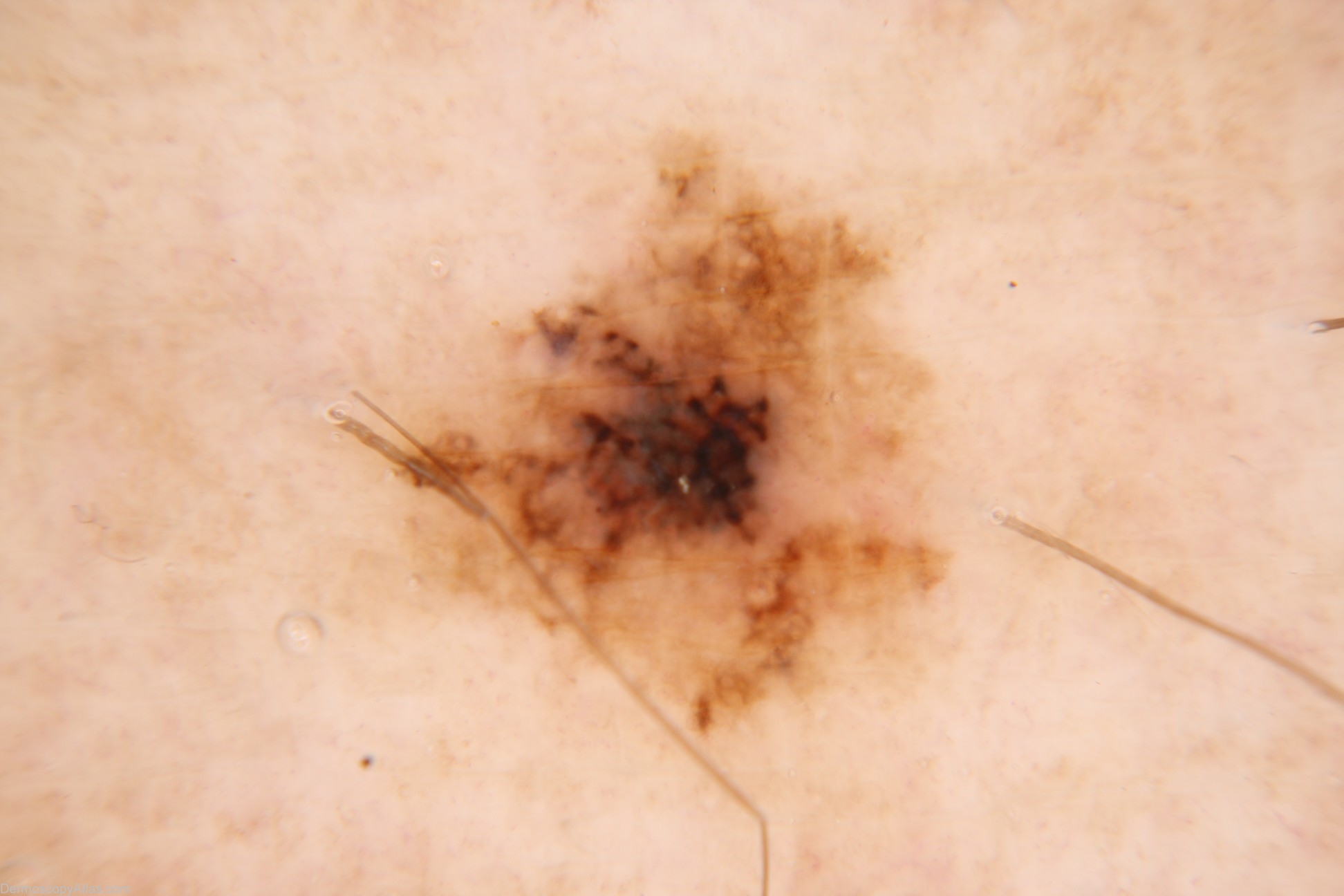
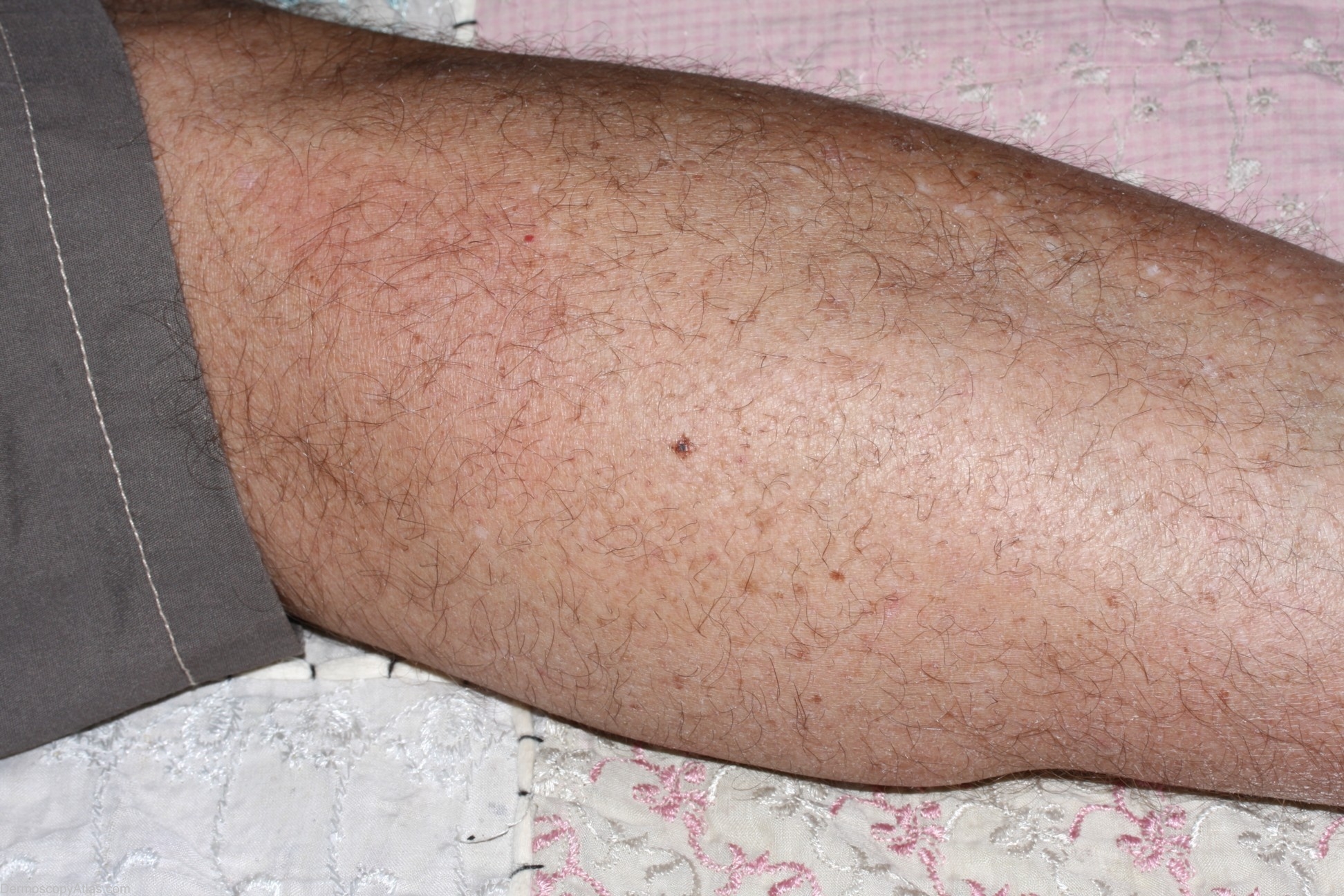
Site: Calf
Diagnosis: Melanoma in situ
Sex: M
Age: 55
Type: Dermlite Non Polarised
Submitted By: Cliff Rosendahl
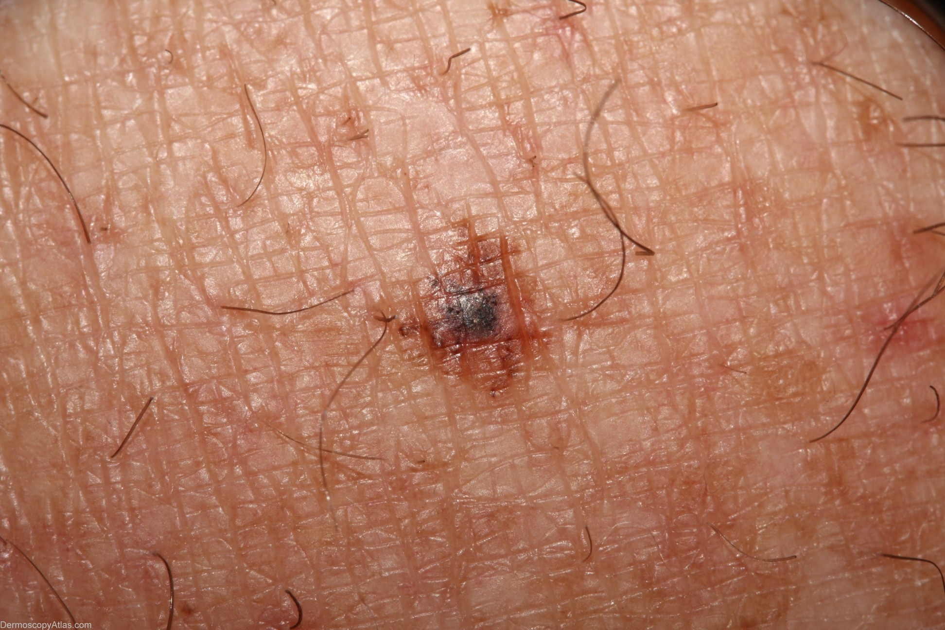
Description: Dermoscopy - This in-situ melanoma exhibits a reticular pigment pattern which is asymmetrical. Colours include tan,black, blue-grey and pink. There are definite pseudopods attached to the broadened pigment network in the central "active" part of the tumour. There are small dot vessels in the pink portion visible in the enlarged view
History: The patient had noticed this as an apparently new lesion and believed that it was changing over a matter of weeks. He had no personal history of skin malignancies but his son had a history of a melanoma at age 20. Histology was of a level 1 melanoma.