Loading
TeleDermConsult-logo
  • Recent Submissions
  • Search
  • Diagnosis
  • Quiz
  • Image comparison
  • Submit
  • Teaching Cases
  • Index
  • About

<< Back to Diagnosis Page

Image Number #2811 (Seborrhoeic keratosis)

Site: Back

Diagnosis: Seborrhoeic keratosis

Sex: M

Age: 65

Type: Dermlite Non Polarised

Submitted By: Alan Cameron

View Full Size Image

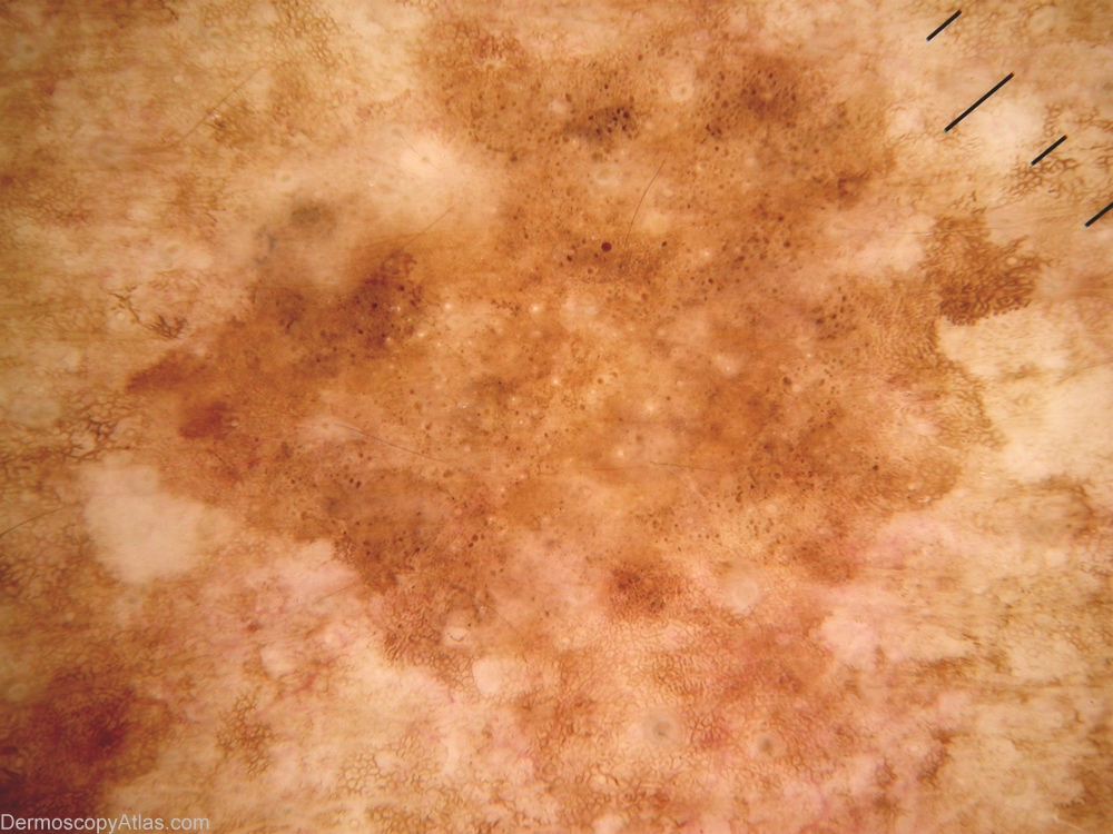
Description: area of lines curved (fingerprinting) far right, a few milia cysts centrally, but extensive brown dots prompted biopsy to exclulde melanocytic lesion.

History: 65yo UK immigrant, bricklayer with extensive occupational sun exposure and actinic damage, multiple previous BCCs. Lesion upper back. Histology reported as reticulate seborrhoeic keratosis with no atypia.

DermNetNZ   eMedicine   PubMed   Dermatology Online   Archives   JAAD for "Seborrhoeic keratosis"

  • Recent Submissions
  • Search
  • Diagnosis
  • Quiz
  • Image comparison
  • Submit an Image
  • Teaching Cases
  • Index
  • About

Copyright 2022. Dermoscopy Atlas. ALL Rights Reserved.

Change Image