
Site: Cheek
Diagnosis: Lentigo Maligna
Sex: F
Age: 74
Type: Dermlite Non Polarised
Submitted By: Ian McColl
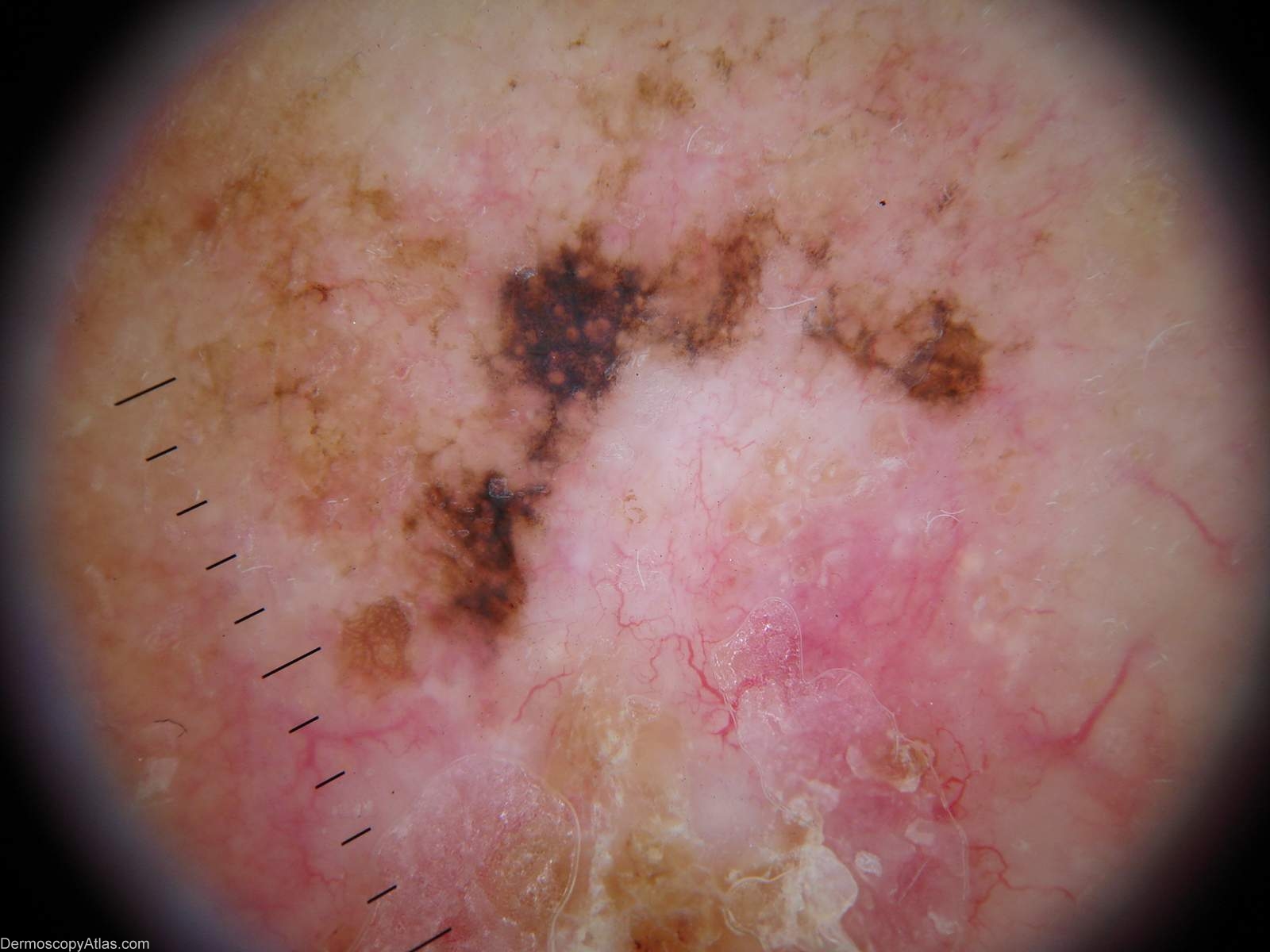
Description: Pigmented lesion on cheek. The dermoscopy shows 2 lesions with an abnormal network, amorphous pigmentation centred around some follicles on the cheek but also the arborising telangiectasia of a BCC and some dermal scaring.
History:
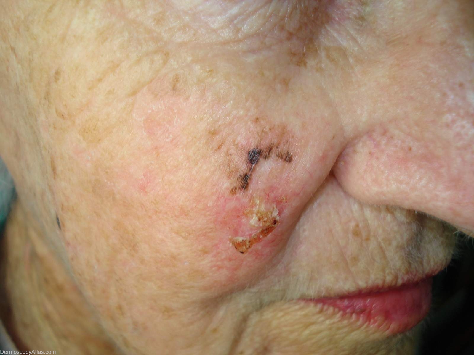
This lady had a lesion removed from her cheek 12 months before by her GP described macroscopically by the pathologist as an ulcerated tan macule 4 by 2mm and reported as an ulcerated BCC transected at a transverse surgical margin. No re excision was done. The patient is legally blind. She subsequently developed this lesion at the excision site.
The pigmented area was reported as a level 1 melanoma and the BCC as a mixed solid and infiltrating. I think from the original pathologist's macro description that she had a lentigo maligna overlying this BCC and both have recurred.
View the Blog discussion of this case.