
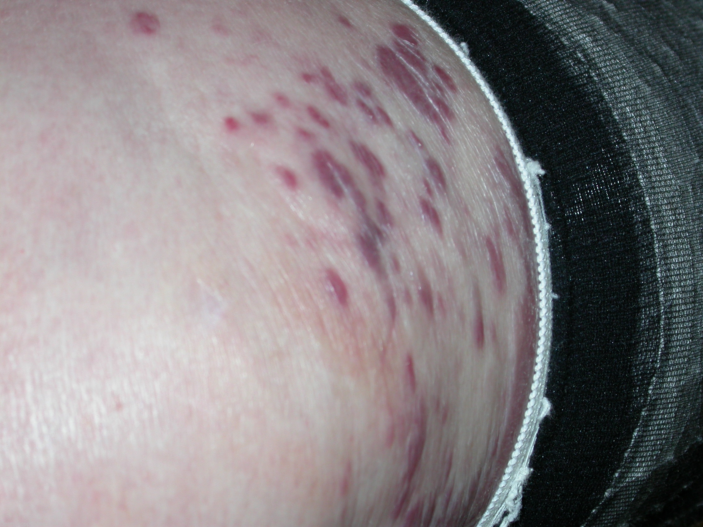
Site: Arms and Legs
Diagnosis: Sarcoma kaposi
Sex: F
Age: 68
Type: Heine
Submitted By: Stelios Minas

Description: Purplish plaques on the thigh.
History:
This elderly woman had slowly progressive confluent purple macules and plaques on her legs associated with increasing edema of her legs for over 5 years. A skin biopsy showed changes typical of Kaposi sarcoma. Histologic sections of skin demonstrate an ill-defined vascular proliferation in the dermis. Higher magnification reveals poorly formed vessels and spindled cells dissecting through the collagen, with focal red blood cell extravasation. The vascular endothelium also demonstrates strong CD31 positivity.These findings are consistent with a dignosis of Kaposi sarcoma.
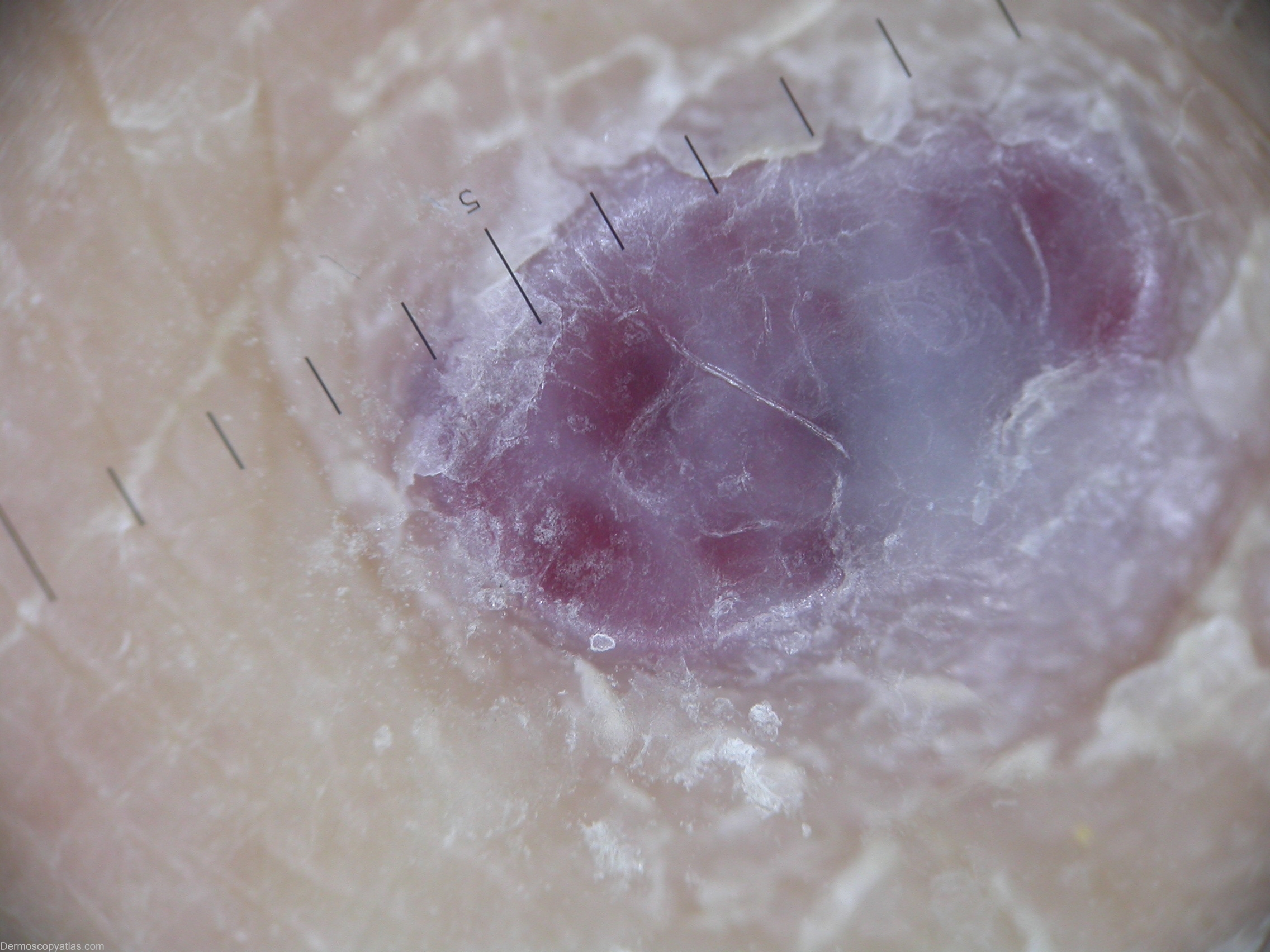
Dermatoscopic description: Bluish and reddish homogeneous structureless. Kaposi sarcoma (KS) was described initially in 1872 by a Hungarian dermatologist, Moritz Kaposi. KS is a spindle-cell tumor thought to be derived from endothelial cell lineage. This condition carries a variable clinical course ranging from minimal mucocutaneous disease to extensive organ involvement. KS can occur in several different clinical settings