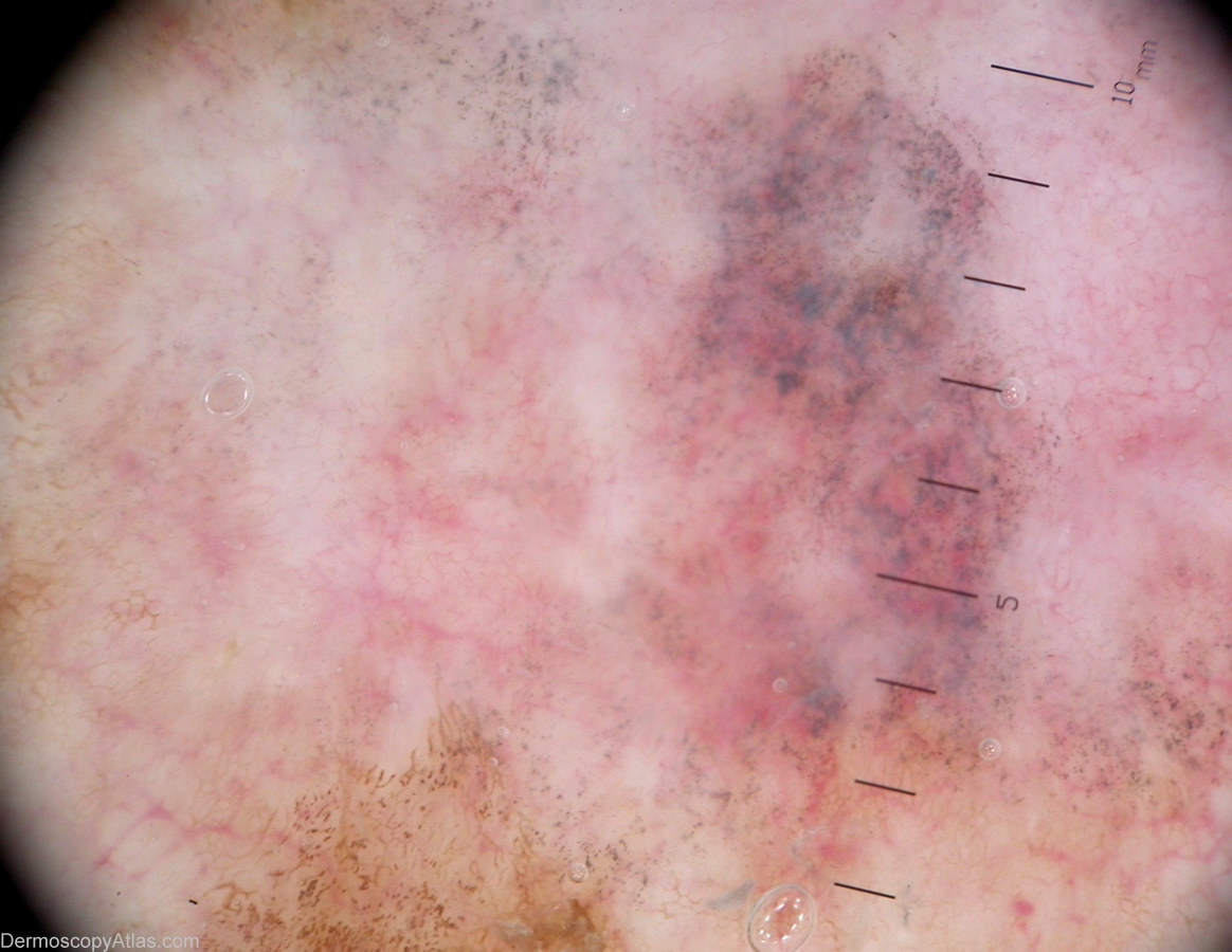
Loading
TeleDermConsult-logo
  • Recent Submissions
  • Search
  • Diagnosis
  • Quiz
  • Image comparison
  • Submit
  • Teaching Cases
  • Index
  • About

<< Back to Diagnosis Page

Image Number #2728 (Melanoma in situ)

Site: Back

Diagnosis: Melanoma in situ

Sex: M

Age: 83

Type: Heine

Submitted By: Alan Cameron

View Full Size Image

Click Here to View Details

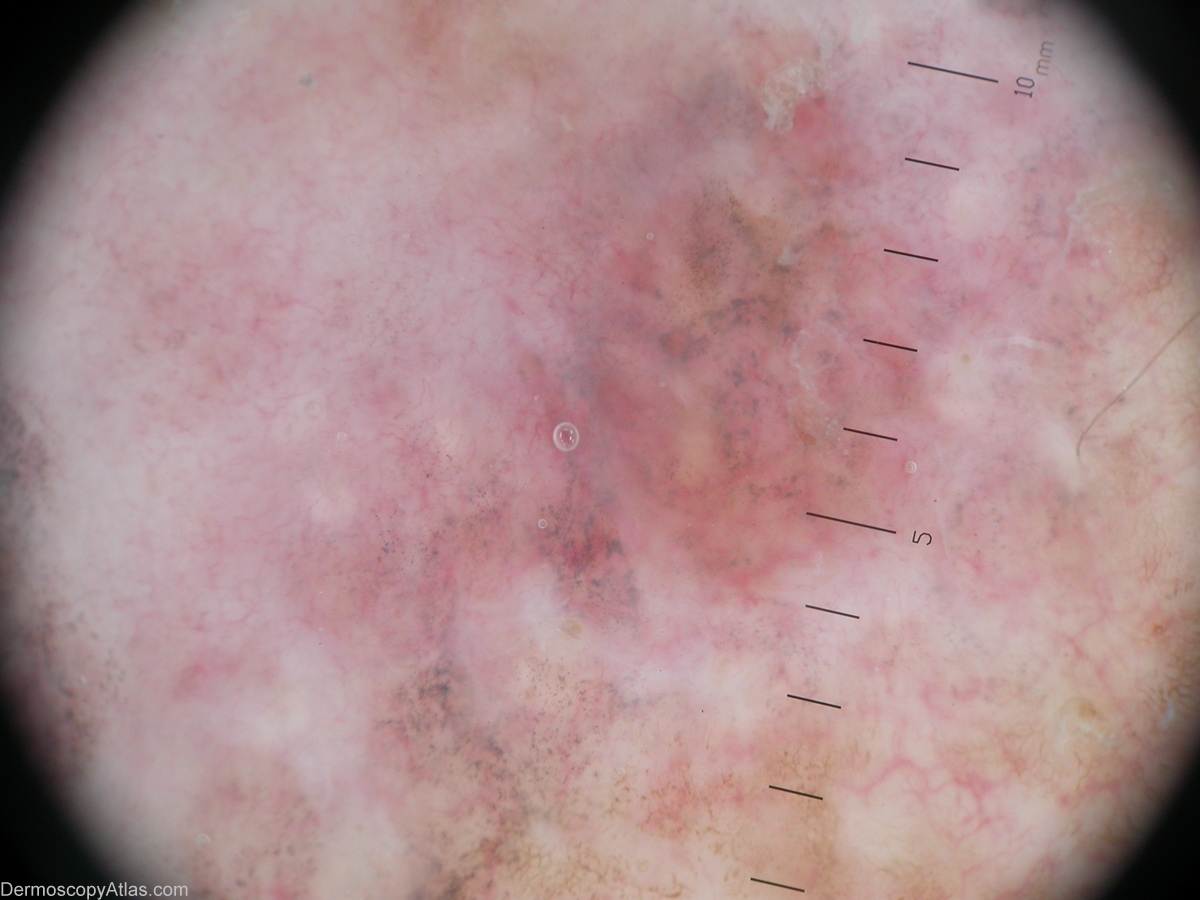
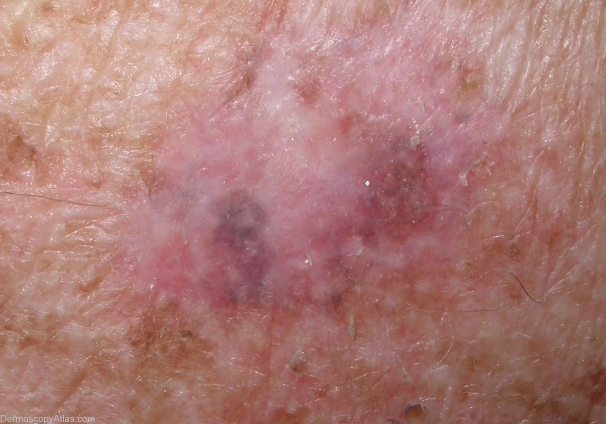
Description: This lesion shows extensive regression with blue/grey dots. No definite melanocytic criteria are noted.

History: Lesion noted on back at skin check, asymptomatic, patient unaware of. PH NMSC only. 2 other melanomas and 2 junctional lentigionous dysplastic naevi also noted on back at this examination. Histology reported as; Sections confirm level 1 (insitu) malignant melanoma with evidence of active and extensive established past regression.

DermNetNZ   eMedicine   PubMed   Dermatology Online   Archives   JAAD for "Melanoma in situ"

  • Recent Submissions
  • Search
  • Diagnosis
  • Quiz
  • Image comparison
  • Submit an Image
  • Teaching Cases
  • Index
  • About

Copyright 2022. Dermoscopy Atlas. ALL Rights Reserved.

Change Image