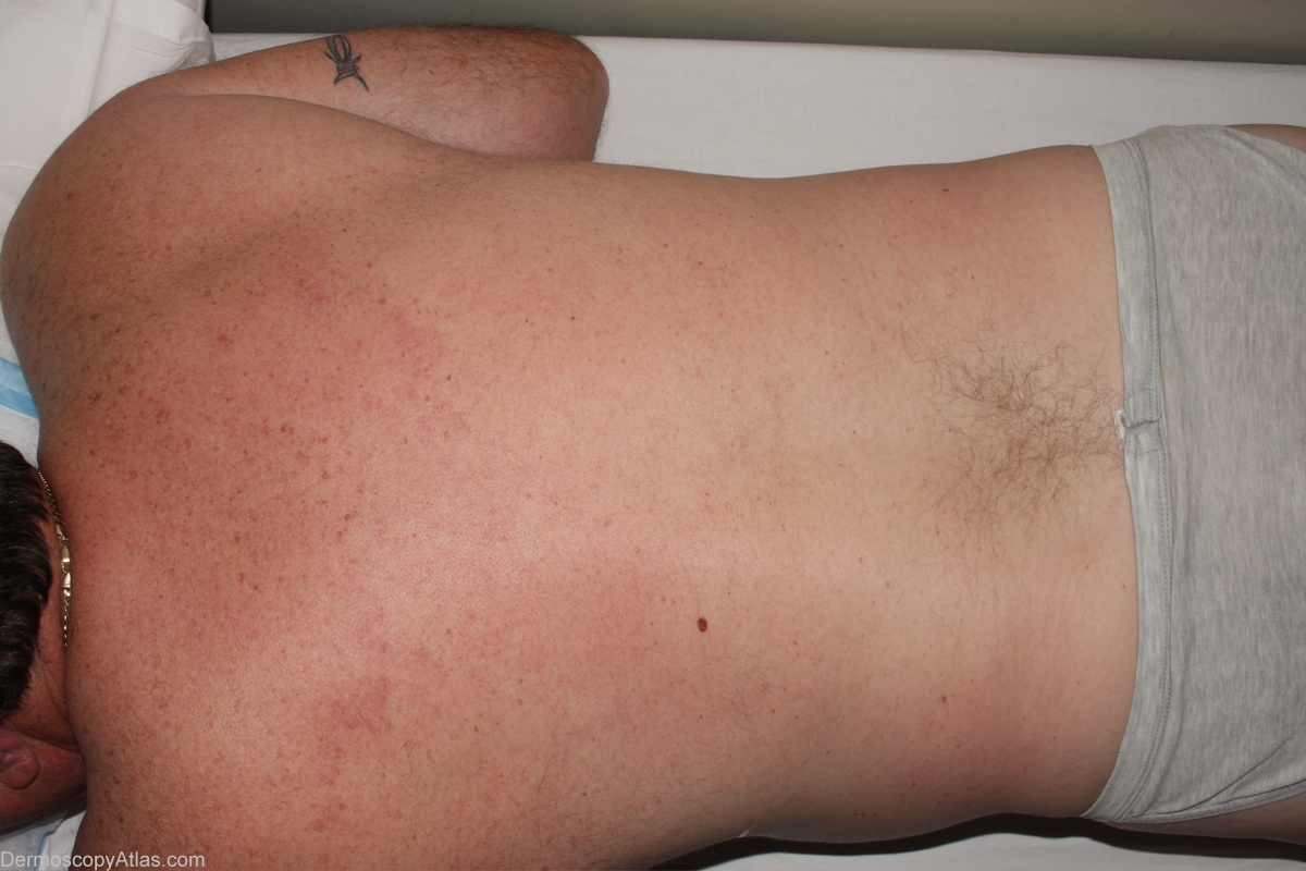
Loading
TeleDermConsult-logo
  • Recent Submissions
  • Search
  • Diagnosis
  • Quiz
  • Image comparison
  • Submit
  • Teaching Cases
  • Index
  • About

<< Back to Diagnosis Page

Image Number #2719 (Nevus dysplastic)

Site: Back

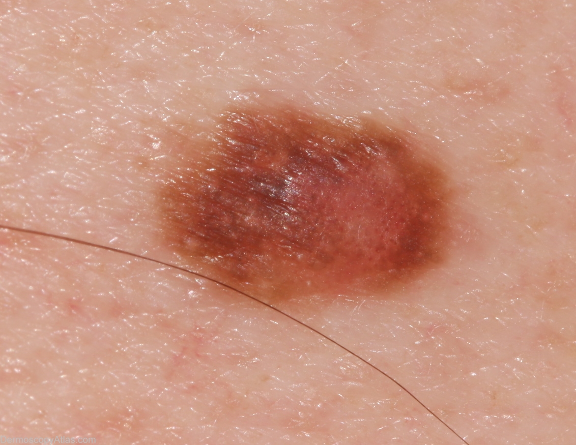
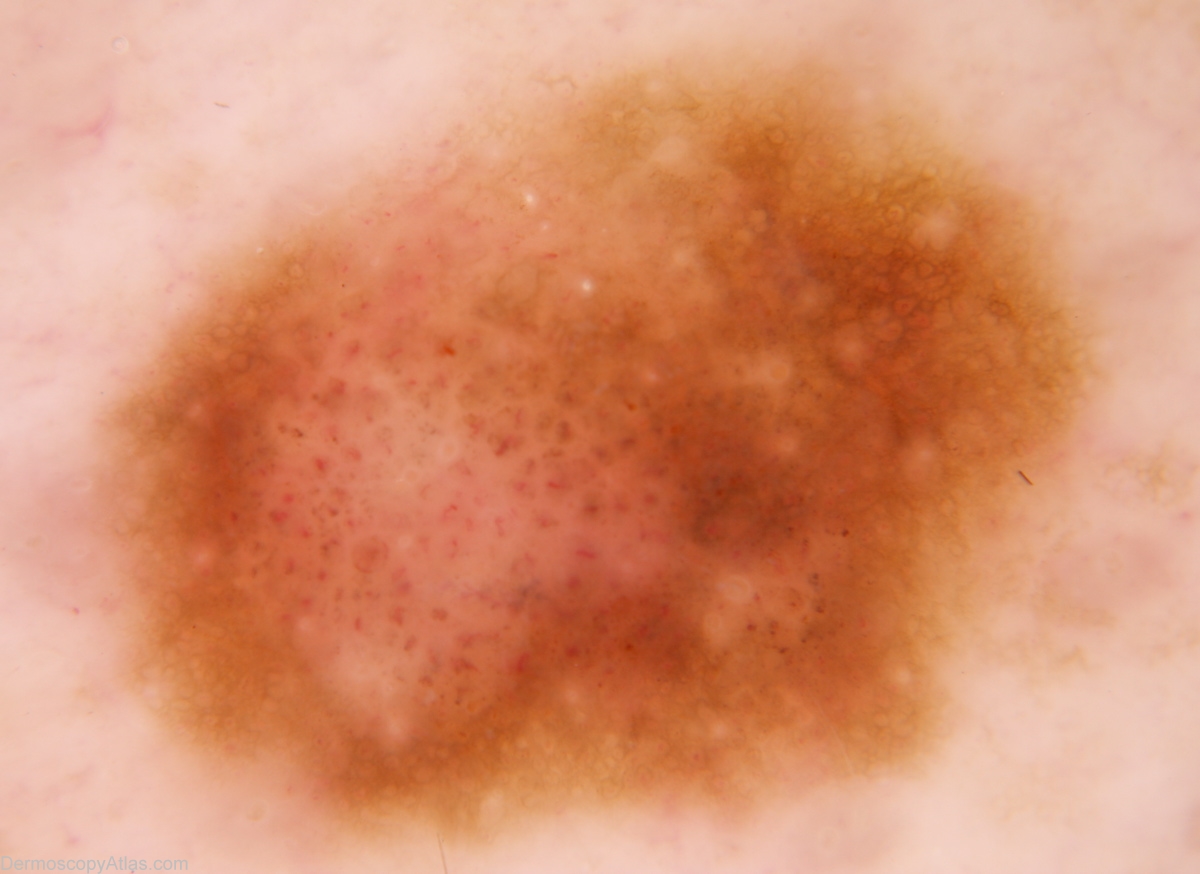
Diagnosis: Nevus dysplastic

Sex: M

Age: 51

Type: Dermlite Non Polarised

Submitted By: Alan Cameron

View Full Size Image

Click Here to View Details

Description:

History: Solitary lesion on back noted to be enlarging over the previous 6 months by wife. No relevant past or family history. Histology reported as; Sections show a dysplastic naevus of early compound type. There is mild to moderate atypia of junctional melanocytes but good maturation of dermal cells. There is no evidence of malignancy.

DermNetNZ   eMedicine   PubMed   Dermatology Online   Archives   JAAD for "Nevus dysplastic"

  • Recent Submissions
  • Search
  • Diagnosis
  • Quiz
  • Image comparison
  • Submit an Image
  • Teaching Cases
  • Index
  • About

Copyright 2022. Dermoscopy Atlas. ALL Rights Reserved.

Change Image