
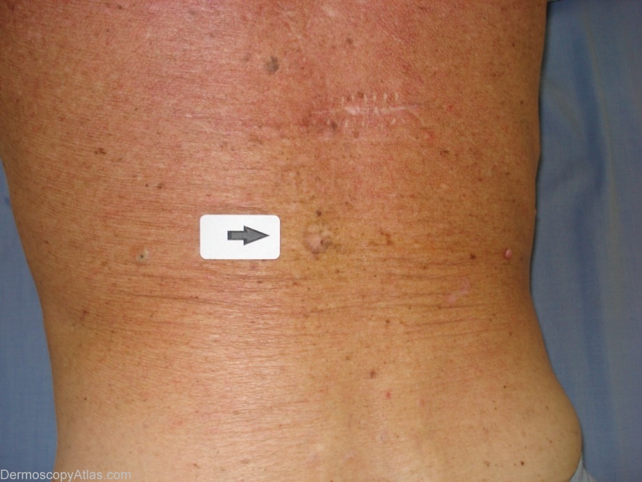
Site: Back
Diagnosis: Pigmented Intraepidermal carcinoma
Sex: M
Age: 68
Type: Dermlite Non Polarised
Submitted By: Cliff Rosendahl
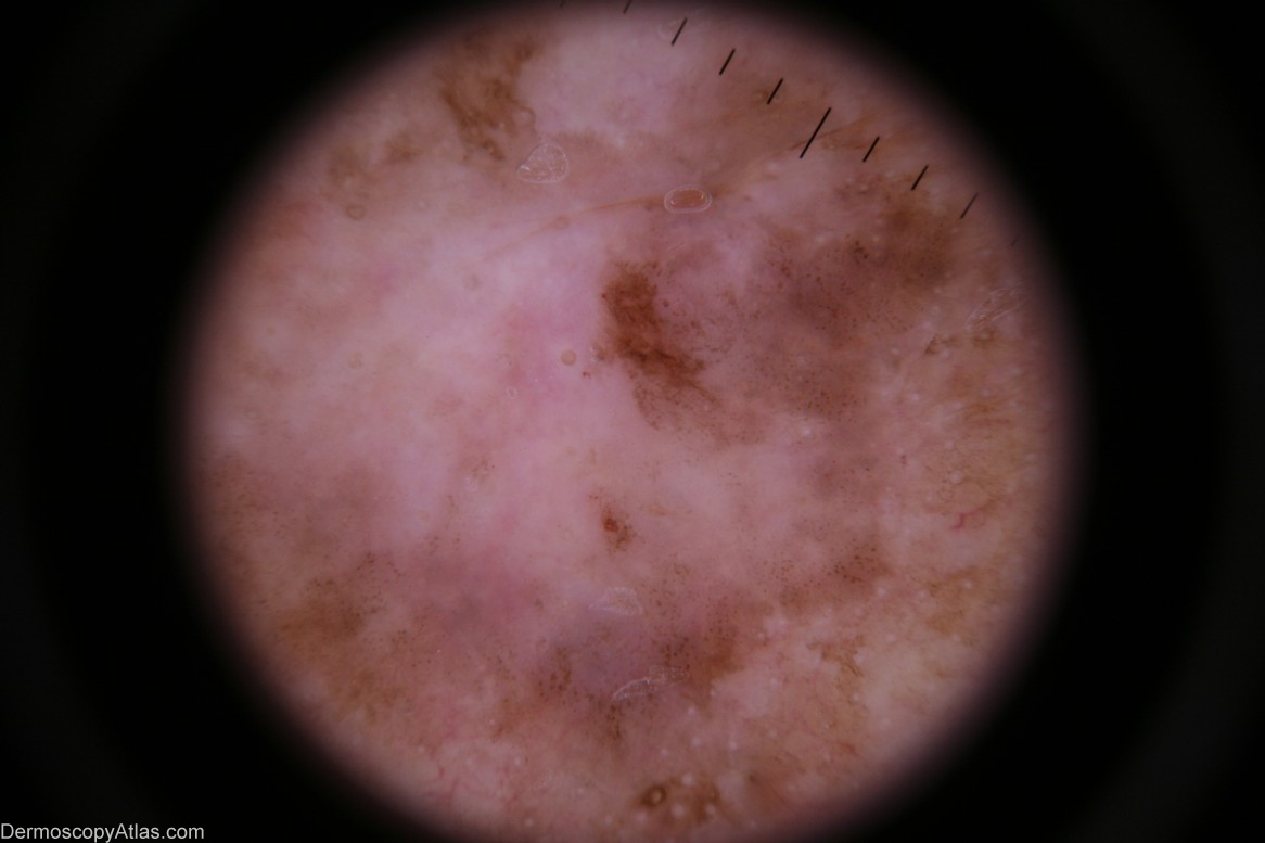
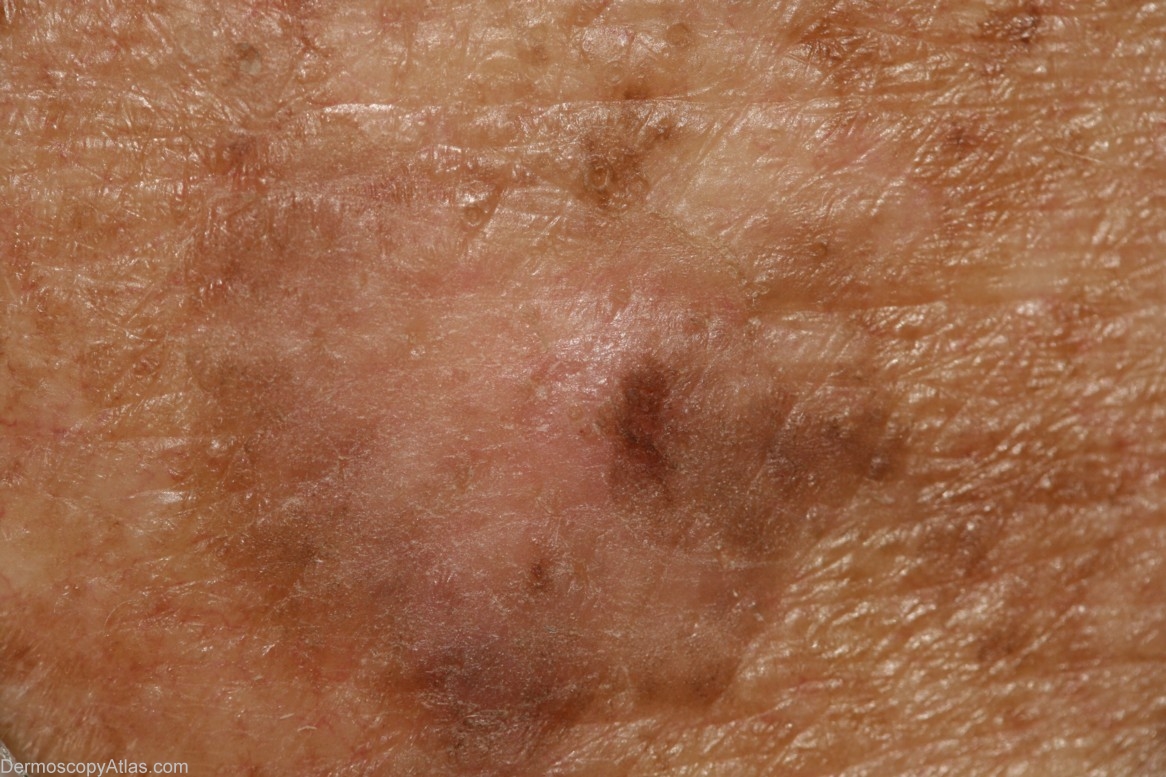
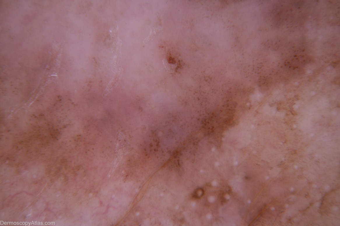
Description: Dermoscopy - There is no pigment network nor are there any other melanocytic features.The distribution of the pigment is asymmetrical. There are milky-red areas and aggregated brown dots. The white circular structures may be milia-like cysts in the adjacent seborrhoeic keratosis which was reported on histology.
History: This lesion was encountered at a routine skin check at the same time as a level 1 melanoma was discovered on his scalp (image 260 in this atlas). Shave biopsy was reported as "...pigmented intraepidermal carcinoma". The definitive excision was reported as "...pigmented intraepidermal carcinoma and seborrhoeic keratosis". IEC,s do sometimes arise in pre-existing seborrhoeic keratoses. This may be the case here or the two lesions may be separate and in collision. The skin on this man's back was severely sun-damaged.