
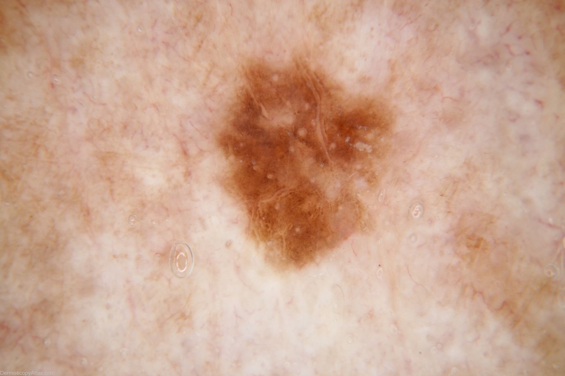
Site: Neck side
Diagnosis: Pigmented Intraepidermal carcinoma
Sex: F
Age: 80
Type: Dermlite Non Polarised
Submitted By: Cliff Rosendahl
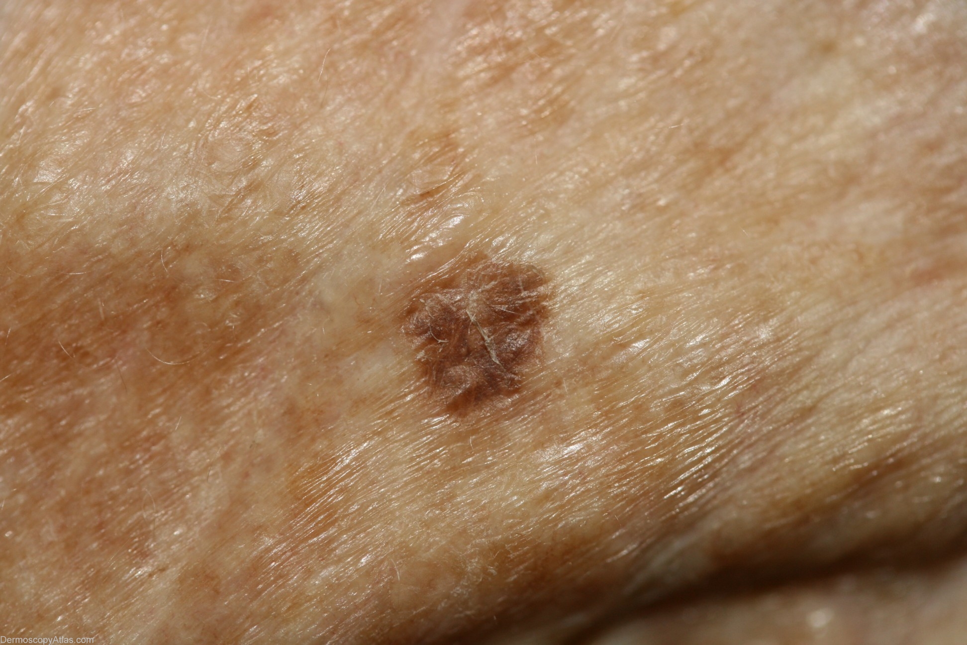
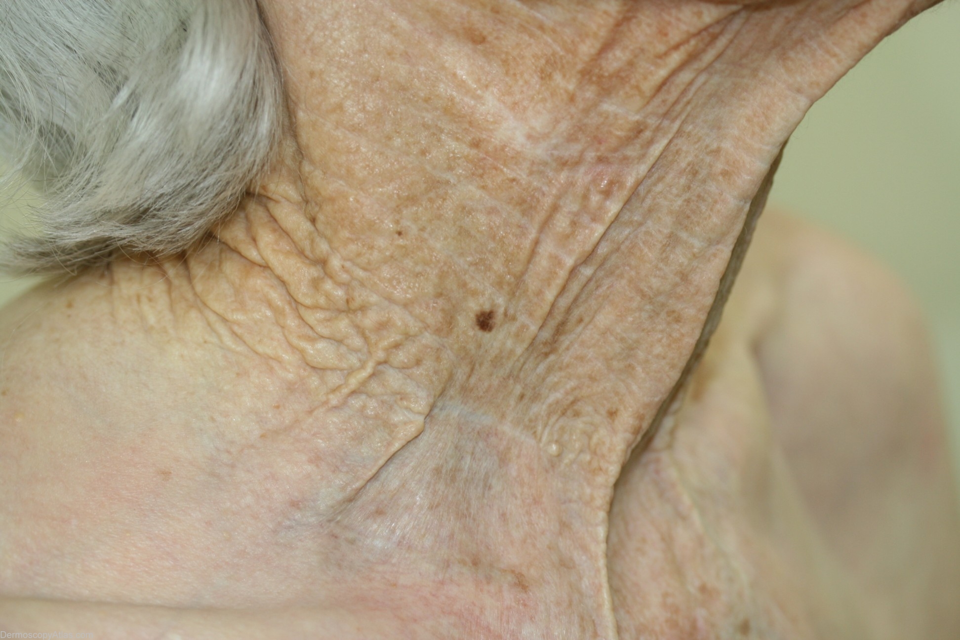
Description: Dermoscopy image - There is no reticular network or pseodonetwork nor are there any other melanocytic features. The white round structures do not have the "glow" of milia-like cysts and they are consistent with follicular openings. There are white lines curved and one area of subtle grey. There are also focal aggregations of brown dots best appreciated in the full-sized view.The main differential diagnosis was seborrhoeic keratosis or solar lentigo with regression explaining the grey. It was reported histologically as simply a pigmented IEC. There was no mention of regression.
History: This small pigmented IEC was encountered at a routine skin check.