
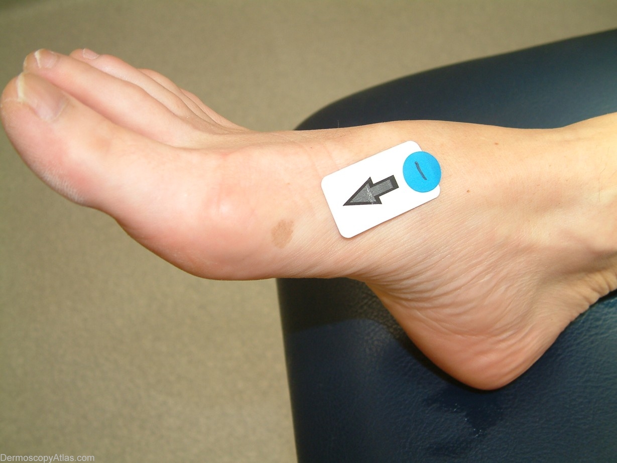
Site: Foot,dorsum
Diagnosis: Tinea nigra
Sex: F
Age: 43
Type: Dermlite Non Polarised
Submitted By: Cliff Rosendahl
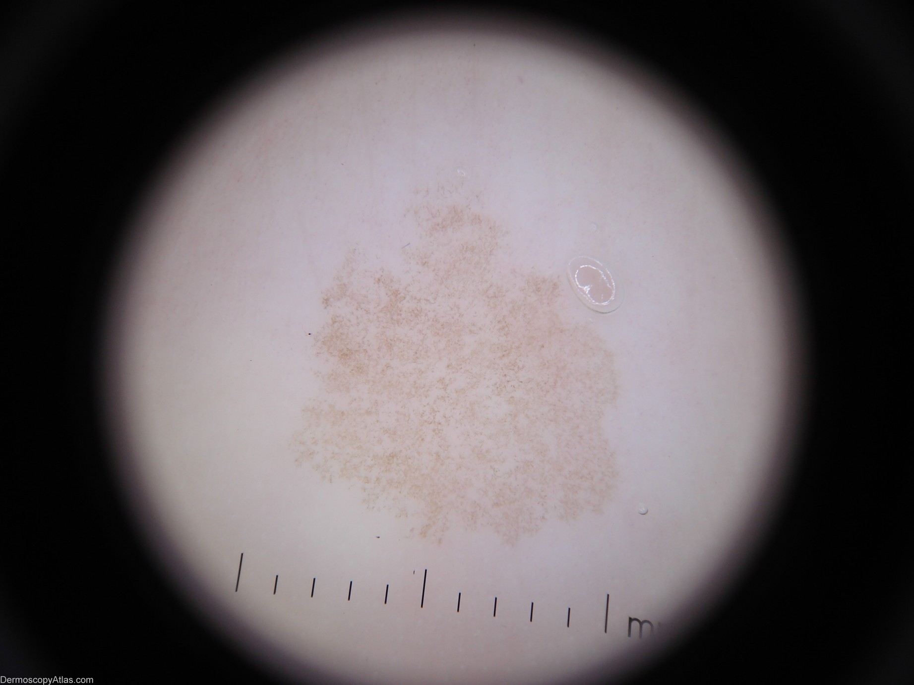
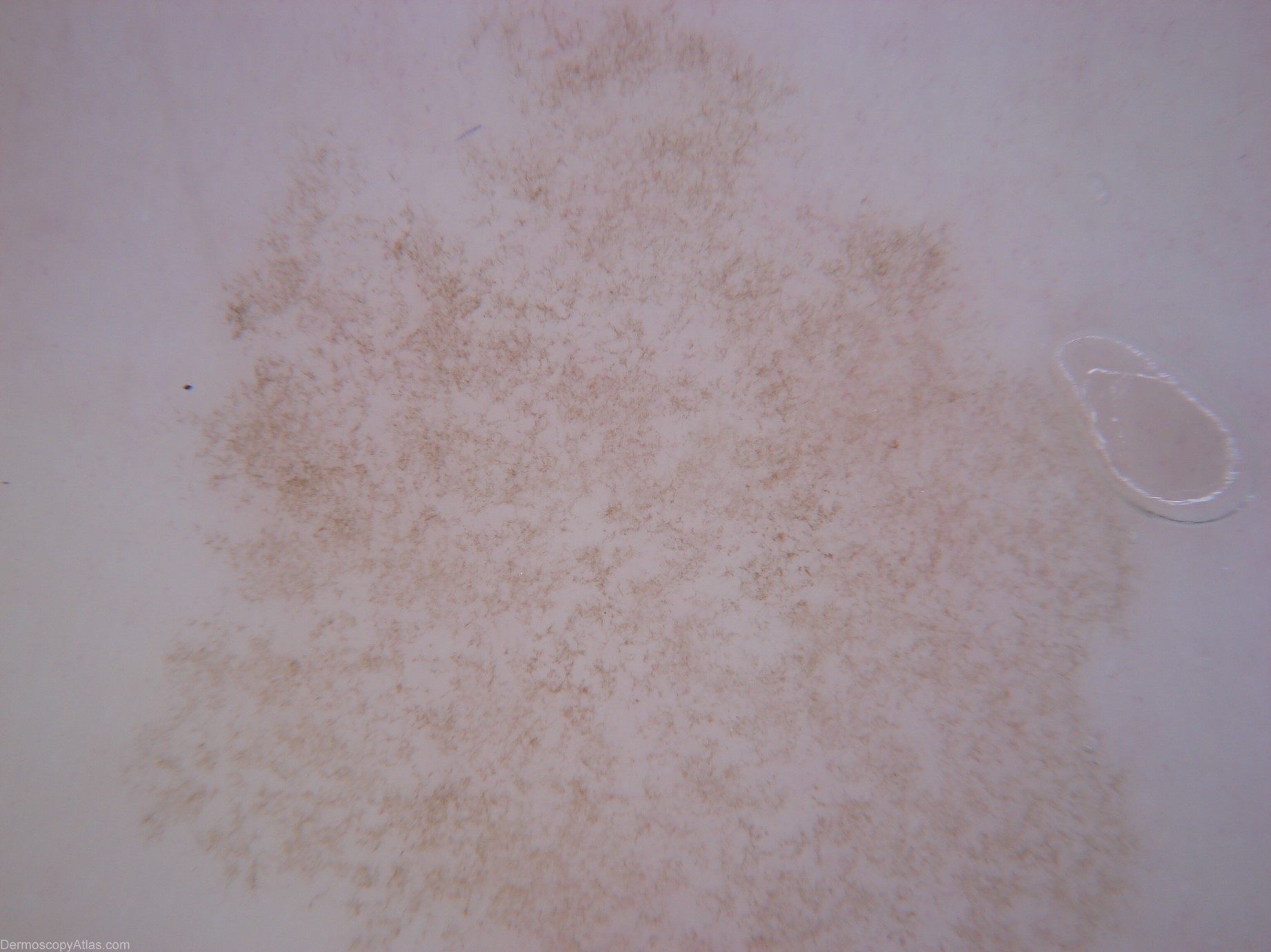
Description: Dermoscopy - There is one colour. Pigment pattern is symmetrical. The pigment appearance is "soft" like a thin veneer of frost on glass. There is no reticular network. The Pseudo-network present is produced by branching hyphae in the stratum corneum of the epidermis. It has neither lines-reticular nor lines-curved. The adjective I would use is "crystalline".
History:
This 42 year old lady with a past history of melanoma presented for a routine skin check and this lesion was encountered
This condition is caused by Exophiala werneckii. It is most commonly found on the palms or palmar surfaces of the fingers and less commonly the plantar or lateral surfaces of the feet. ( Ref Skin Pathology - Weedon 2nd edition page 674)