
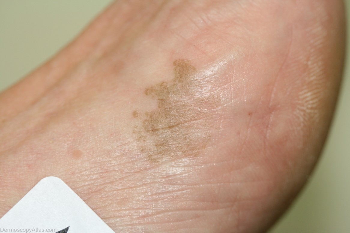
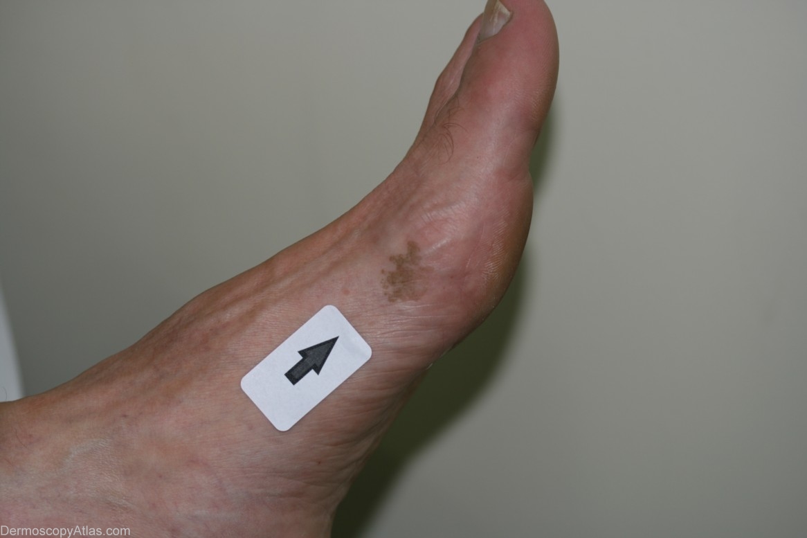
Site: Foot,dorsum
Diagnosis: Tinea nigra
Sex: M
Age: 59
Type: Dermlite Non Polarised
Submitted By: Cliff Rosendahl
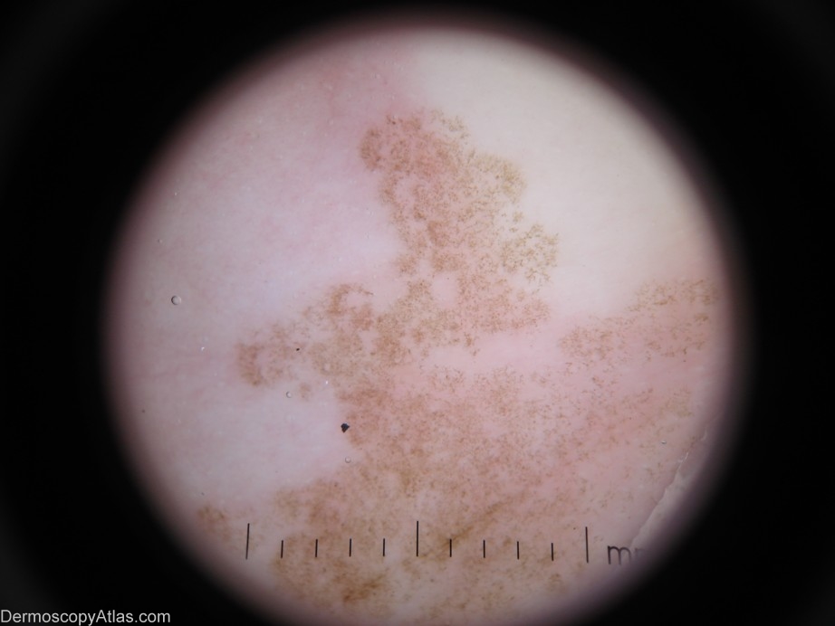
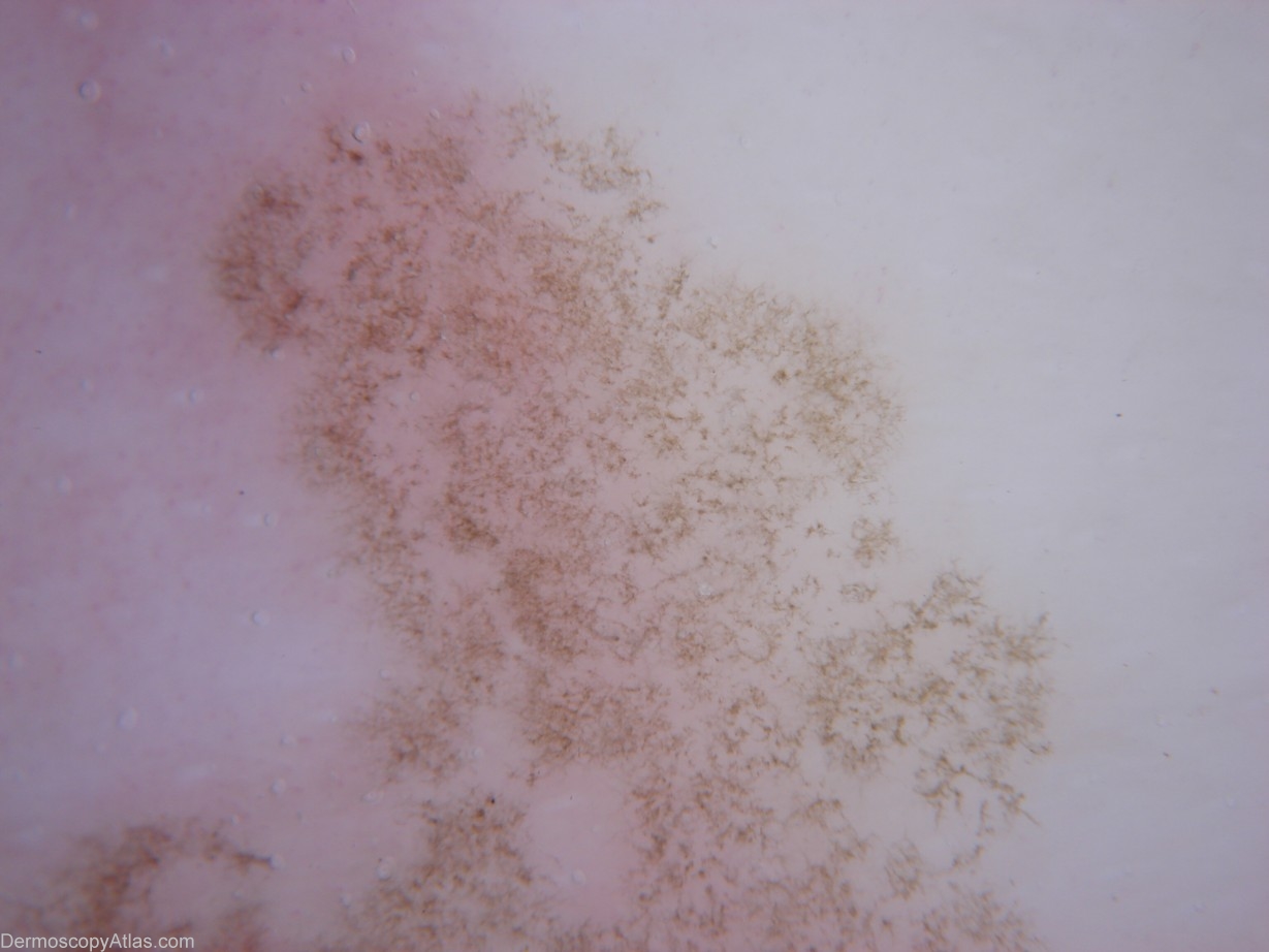
Description: Dermoscopy - There is only one colour present. The pattern is symetrical (but not the shape). The pattern is also very "soft" and reminds me of a thin veneer of frost (which is about to melt)on glass. The Pseudo-network present is produced by branching hyphae in the stratum corneum of the epidermis. It has neither lines-reticular nor lines-curved. The adjective I would use is "crystalline".
History: This man presented with a pigmented macule which had been noticed to be present for 2 years and to be slowly enlarging. This condition is caused by Exophiala werneckii. It is most commonly found on the palms or palmar surfaces of the fingers and less commonly the plantar or lateral surfaces of the feet. ( Ref Skin Pathology - Weedon 2nd edition page 674)