
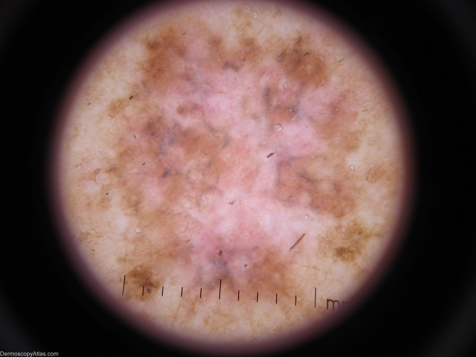
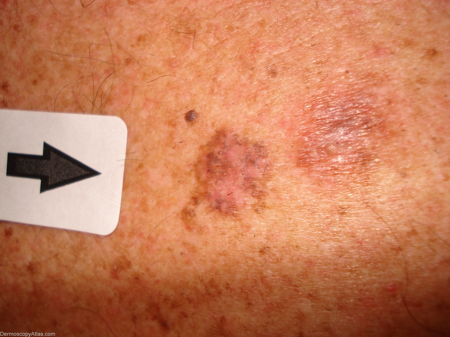
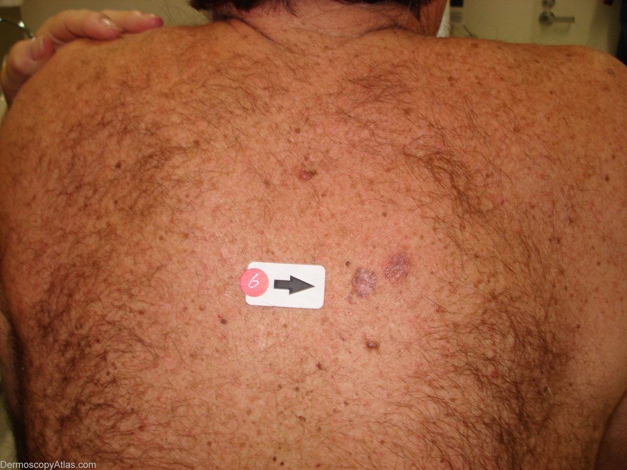
Site: Back
Diagnosis: Melanoma invasive
Sex: M
Age: 57
Type: Heine
Submitted By: Cliff Rosendahl
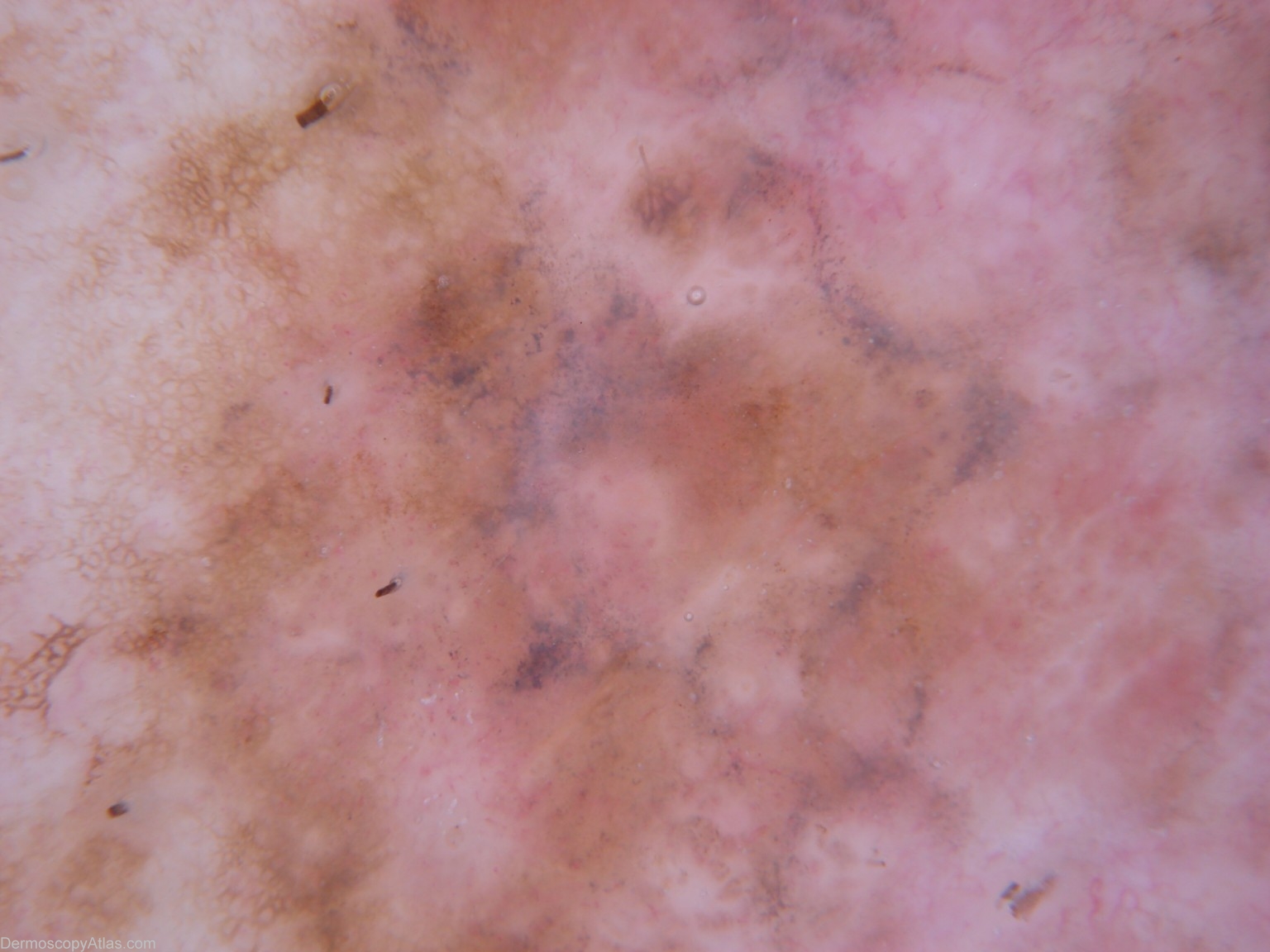
Description: This image demonstrates multiple colours including the blue-grey dots due to regression. The lesion shows a quite disordered pattern with fragments of reticular pigment network interspersed with more structureless areas
History: This 57 year old gentleman had been referred to a plastic surgery clinic for definitive re-excision of a level 1 melanoma from his costal margin. I was asked to examine him for the presence of any other significant lesions. This lesion on his back was subjected to excision biopsy and histology revealed a level 2 superficial spreading melanoma arising in a lentiginous junctional dysplastic naevus. Breslow thickness was 0.4 mm.