
Site: Back
Diagnosis: Melanoma invasive
Sex: M
Age: 51
Type: Heine
Submitted By: Jean-Yves Gourhant
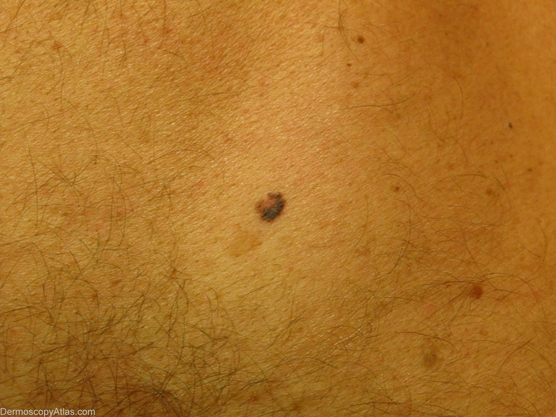
Description: Clinical view: a suspicious pigmented lesion.
History: A 51 years old patient whose wife had noticed this small asymmetric pigmented lesion of the upper back.
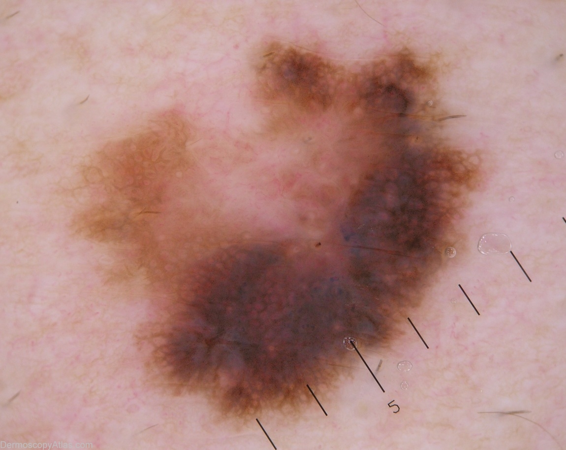
Dermoscopy shows asymmetry, irregular network, irregular streaks, irregular black and grey dots, a blue white veil. The white central patch has the same colour than the normal skin, and is not regression. It exhibits some linear irregular vessels
The pathology reported a superficial spreading melanoma, Clark III, Breslow 0.46, without any regression.