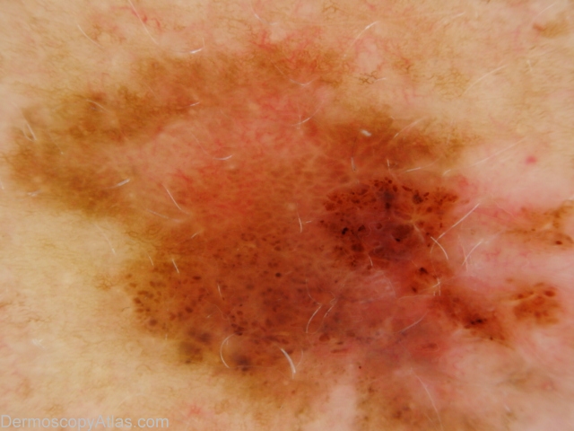
Loading
TeleDermConsult-logo
  • Recent Submissions
  • Search
  • Diagnosis
  • Quiz
  • Image comparison
  • Submit
  • Teaching Cases
  • Index
  • About

<< Back to Diagnosis Page

Image Number #2615 (Melanoma invasive)

Site: Abdomen

Diagnosis: Melanoma invasive

Sex: F

Age: 54

Type: Dermlite Polarised

Submitted By: Ian McColl

View Full Size Image

Click Here to View Details

Description: Pigmented lesion on the back showing pigment dots, asymmetry of pigmentation and a negative network.

History: This lesion had developed over several months. It was brought to her attention by being itchy. It was a superficial spreading melanoma, 0.35mm thick , Clark level 2. The histopathology did show some features of regression.

DermNetNZ   eMedicine   PubMed   Dermatology Online   Archives   JAAD for "Melanoma invasive"

  • Recent Submissions
  • Search
  • Diagnosis
  • Quiz
  • Image comparison
  • Submit an Image
  • Teaching Cases
  • Index
  • About

Copyright 2022. Dermoscopy Atlas. ALL Rights Reserved.

Change Image