
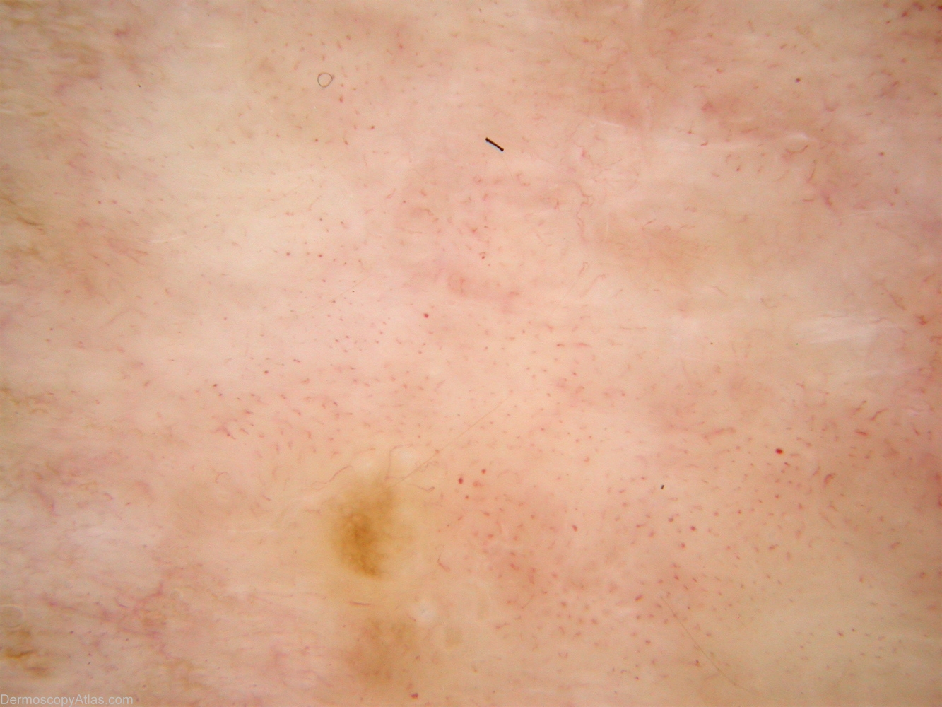
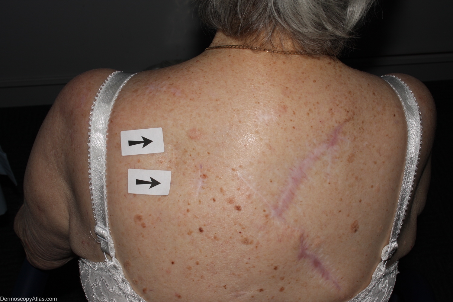
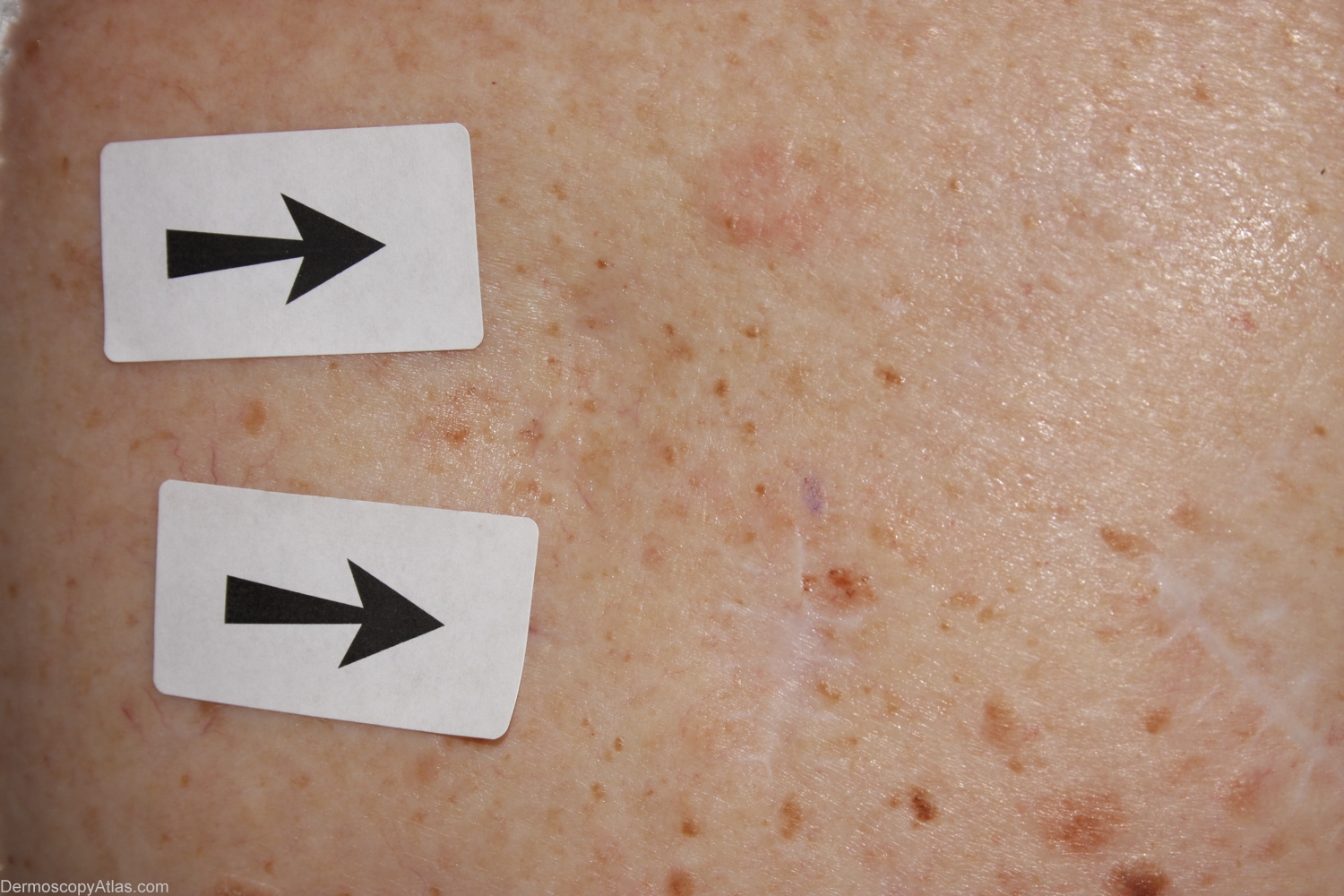
Site: Back
Diagnosis: Melanoma amelanotic
Sex: F
Age: 79
Type: Mixed
Submitted By: Alan Cameron
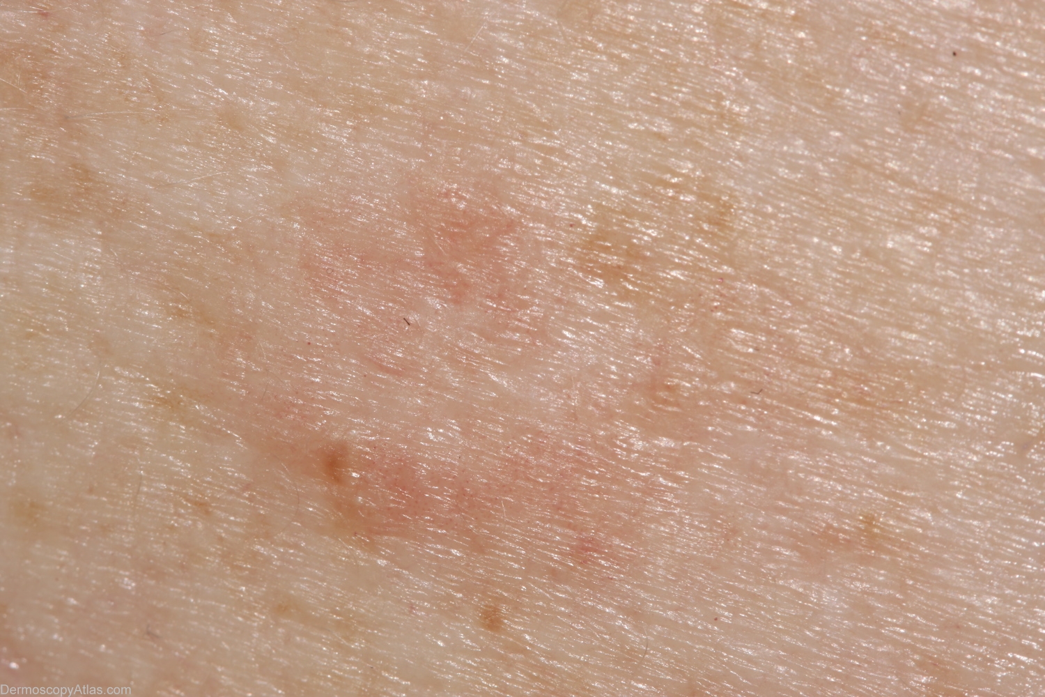
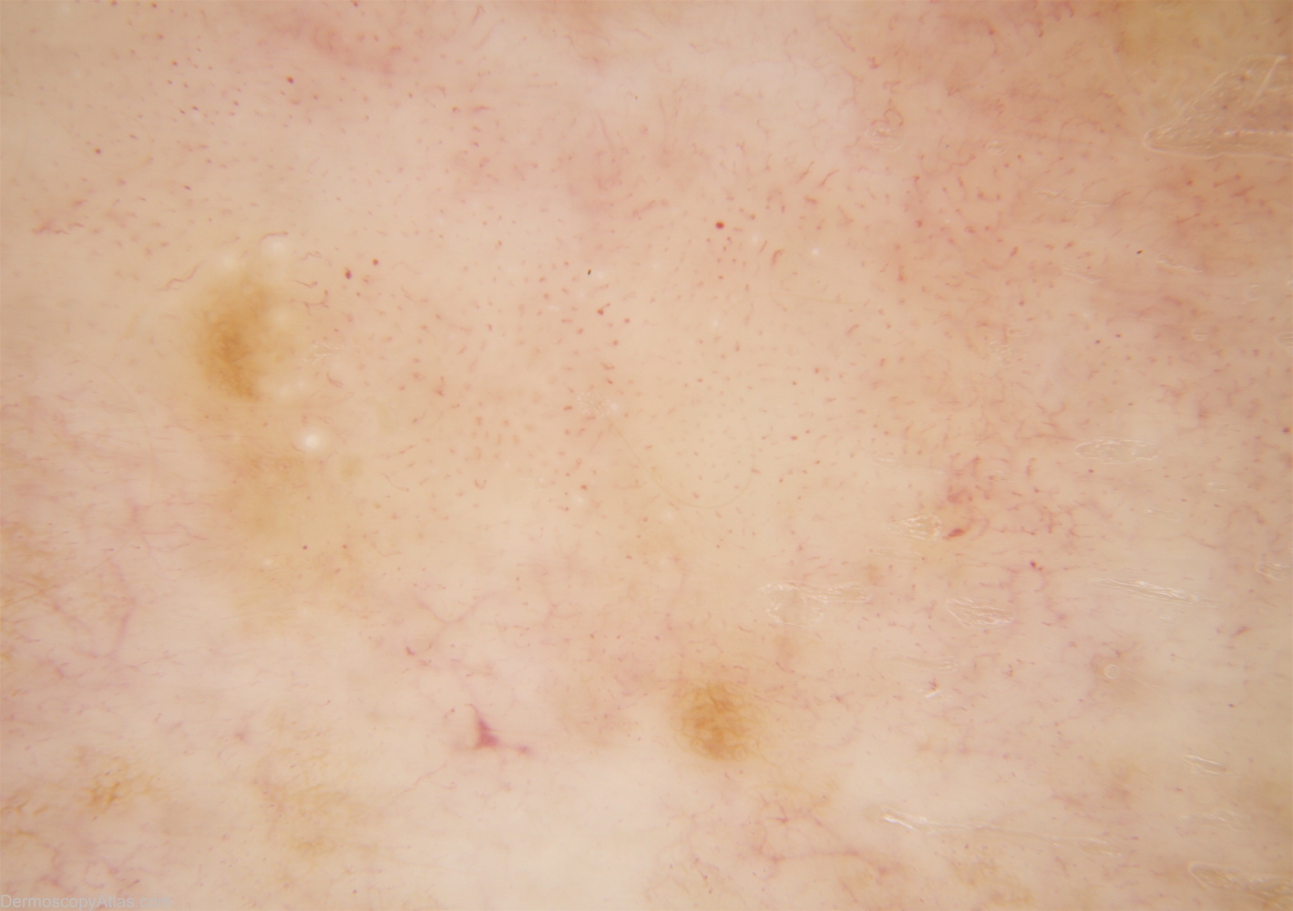
Description: Contact Polarised Dermlite IIHR Vessels are polymorphous. Linear irregular at 7 o'clock, irregular thin hairpin at 3 and 9, comma and dot centrally. Shiny white structures top right are polarisation artefact.
History: This 79 year old woman has had 9 previous melanomas. Two lesions marked for biopsy, lesion in question superior. Histology was reported as — Sections show a few nests of superficial basal cell carcinoma admixed with a very focal level 2 (0.3mm thick) lentigo maligna melanoma. It is arising in a larger in situ lesion. There is no ulceration or dermal mitoses. There is a mild lymphocytic infiltrate but little regression.