
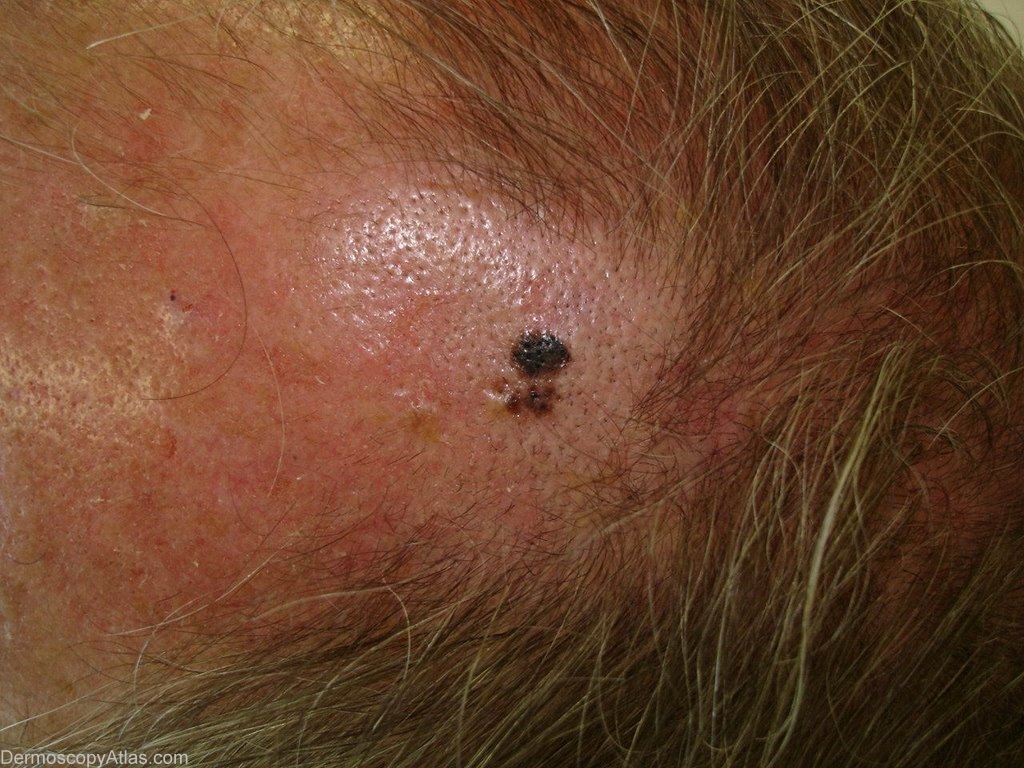
Site: Scalp
Diagnosis: Melanoma superficial spreading
Sex: M
Age: 65
Type: Dermlite Polarised
Submitted By: Ian McColl
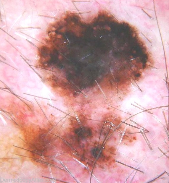
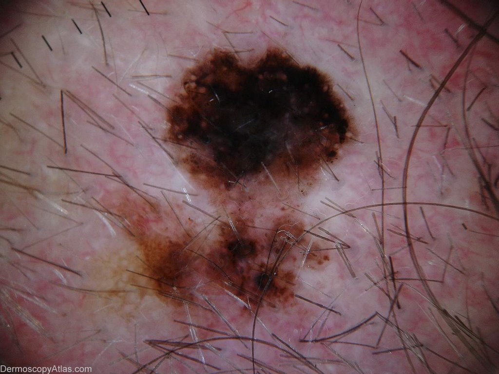
Description: Dermatoscope image On the dermoscopy image, I feel there there is a network, so the lesion is melanocytic. It has asymmetrical pigmentation and several features of melanoma such as: Blue-white veil, Broadened network (10 o'clock), Radial streaming (2 o'clock), Multiple colours, Possibly peripheral black dots/globules
History: He had only noticed this over the last 6 months.It was not his only presenting problem.I have shaved his hair to photograph it.I noted it when examining him and thought it was a dark seb k but then put the dermatoscope to it and changed my mind!
It was reported as a level 2 0.4mm superficial spreading melanoma.
I think there has been some regression in the centre.
The dermatoscope picture shows a dense pigment pattern and a blue grey veil with radial streaming and peripheral dots and globules.
Dark looking seborrhoeic keratoses always merit a quick look with the dermatoscope especially if flat.
View the Blog discussion of this case.