
Site: Back
Diagnosis: Melanoma superficial spreading
Sex: M
Age: 72
Type: Molemax
Submitted By: Wynn Hlaing
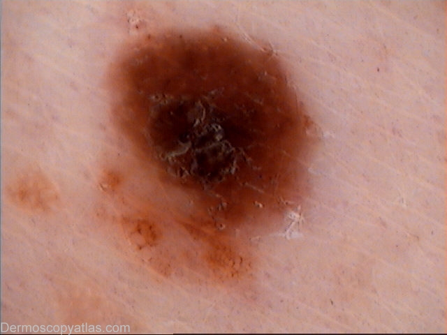
Description: Dermoscopy-'Fried egg'- appearing brown macule.
History:
This 'fried egg' appearing pigmented lesion on the right mid lumbar area was excised .There is scaly,black lamella in the centre with reticular pigment remnant in the inferior part of the lesion.
Histopathology: MELANOMA Clark Level II with Breslow thickness of 0.7 mm.It is a superficial spreading melanoma. There is mild solar elastosis and associated dysplastic junctional naevus noted.
Differential diagnosis of this lesion includes: Clark(dysplastic) naevus,combined naevus and melanoma.
Black lamella represents superficial pigmented parakeratotic cells.