
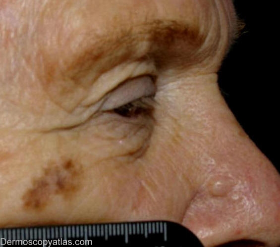
Site: Cheek
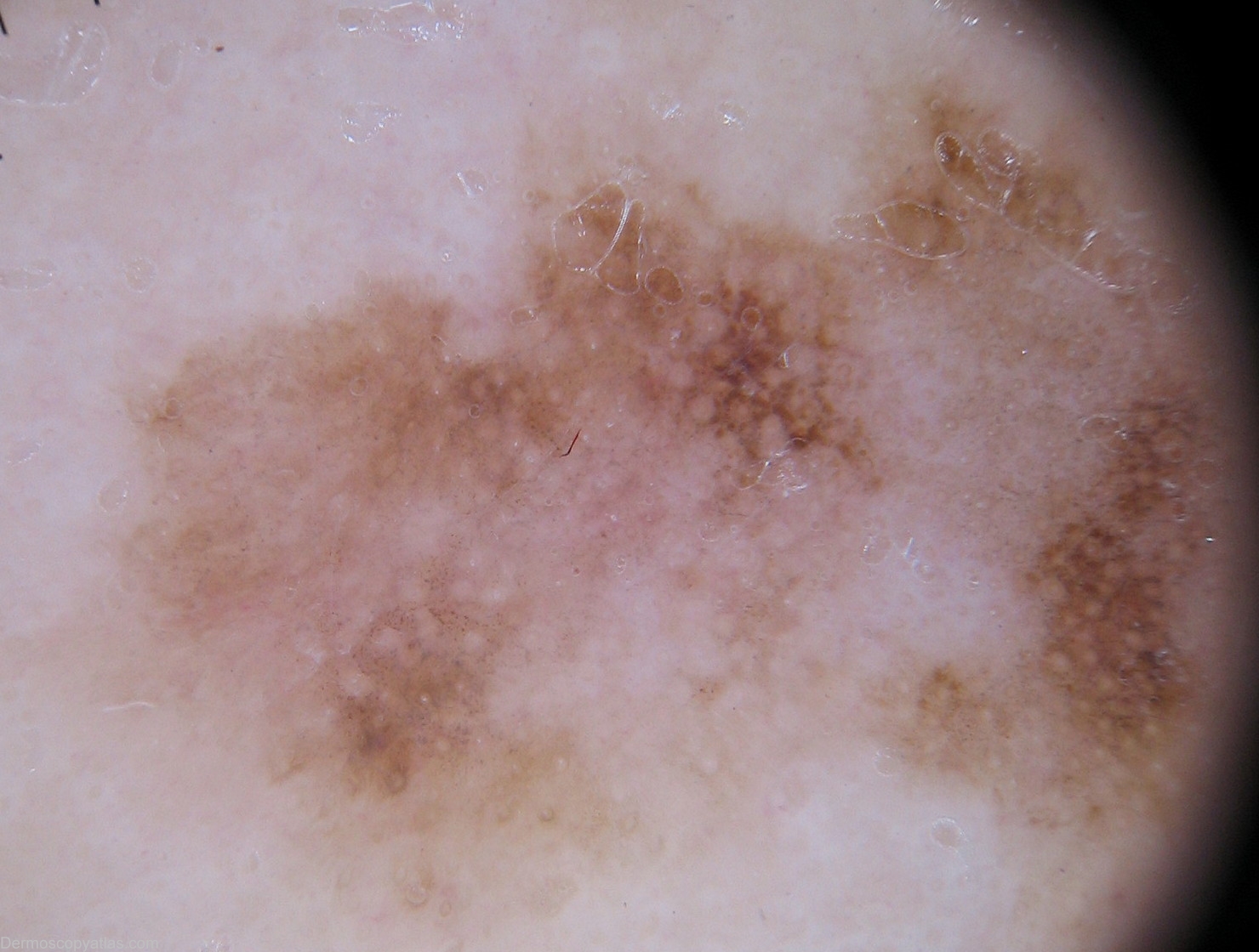
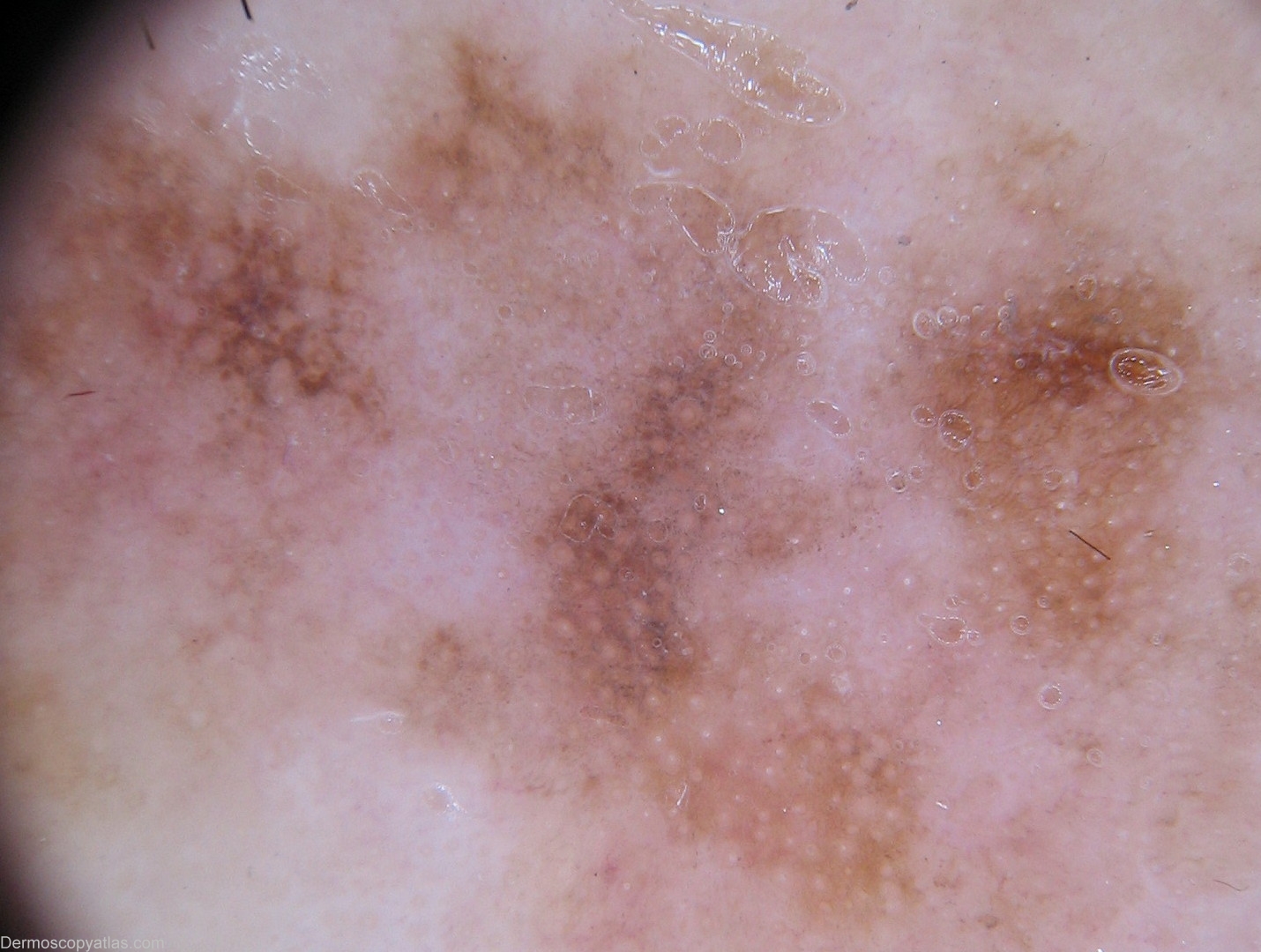
Diagnosis: Lentigo Maligna
Sex: F
Age: 79
Type: Dermlite Non Polarised
Submitted By: Wynn Hlaing
Description: Clinical- Macule with different shades of brown colour.Dermal naevus on the lateral wall of the nose is also seen here.
History:
She has had longstanding pigmented lesion on her right malar area for more than five years which has been slowly increasing in size and darkening in pigmentation.Her skin phototype is I-II.She has no relevant history of skin cancer.
Histopathology: Areas at least amounting to LENTIGO MELANOMA,CLARK LEVEL I,but with possible focal early LEVEL II,and features of patchy regression.
Schiffner R et al.described the "Progression model of lentigo maligna" and Stolz W also described the four characteristic features of lentigo maligna :
Asymmetric pigmented follicular openings,multiple gray dots and globules,dark rhomboidal structures and homogenous brown to dark areas occluding the follicular opening with an irregular outline.
APFOs forming an annular-granular pattern (Cognetta sign) is quite common in lentigo maligna but rare in pigmented actinic keratosis and lentigo senilis(flat seborrheic keratosis).