
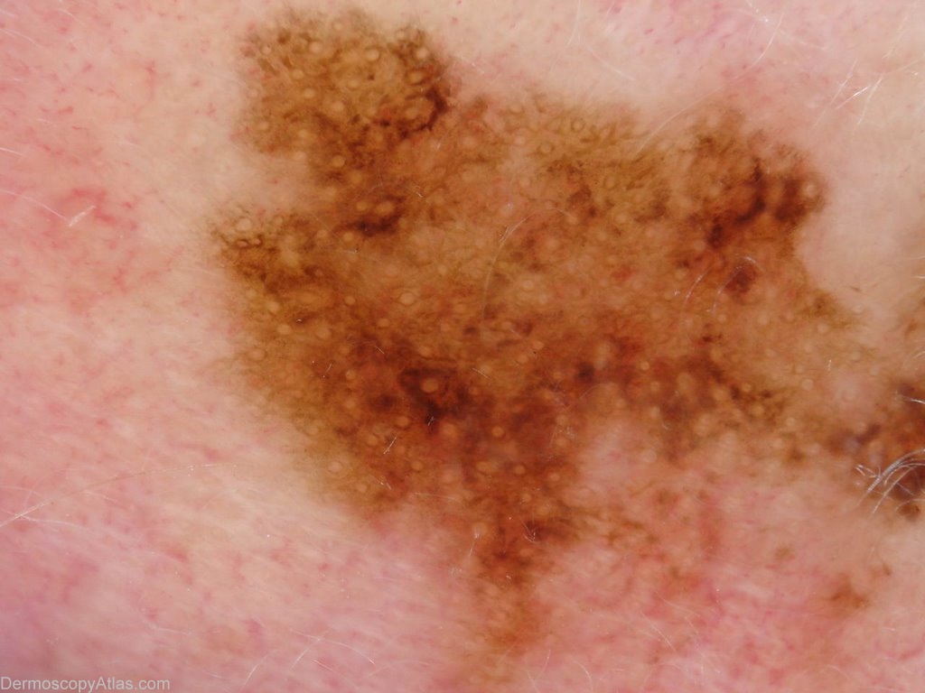
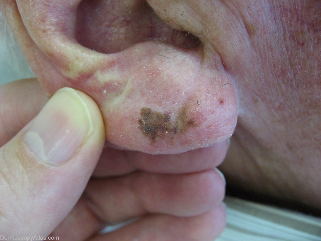
Site: Ear
Diagnosis: Lentigo Maligna
Sex: M
Age: 81
Type: Dermlite Polarised
Submitted By: Ian McColl
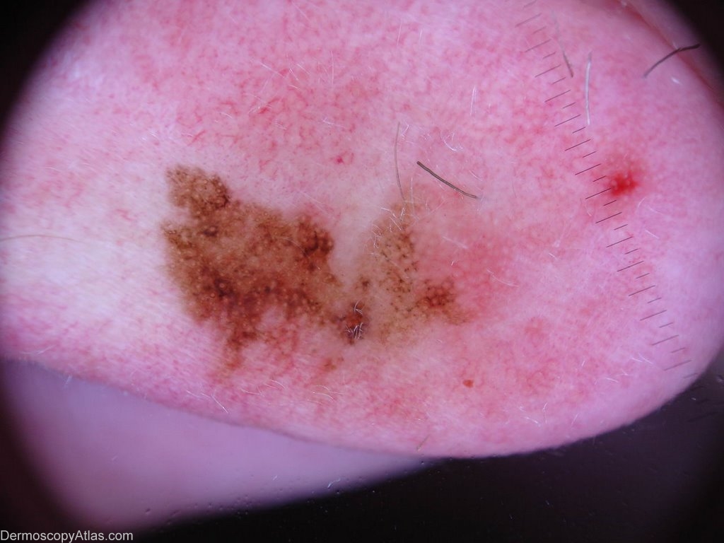
Description: Close up dermatoscope view showing asymmetric pigmented follicular openings and the start of rhomboid structures.
History:
This pigmented lesion on the ear had been slowly growing for some time. Areas had darkened.
The path report was as follows Sections show a shave biopsy of an atypical melanocytic proliferation. For the most part there is a lentiginous proliferation of atypical melanocytes within the basal epidermis. A few nests are present towards one edge. The epidermis is atrophic and the underlying dermis shows marked solar damage. Overall the changes are best regarded as a lentigo maligna (Hutchinson's melanotic freckle). There is no evidence of dermal involvement by the melanocytes. I think the clinical image is all you need to make this diagnosis.
There was a paper recently looking at melanocytic proliferation around these lesions in sun exposed areas. This proliferation is not unusual but the cells have different genetic characteristics to those comprising the lentigo maligna. The inference was that you can over estimate the excision margins you need for these lesions. The proof of the pudding are the number who develope recurrence of lentigo maligna if you do not take them into consideration!
View the Blog discussion of this case.