
Site: Arm,upper
Diagnosis: Melanoma superficial spreading
Sex: F
Age: 33
Type: Molemax
Submitted By: Wynn Hlaing
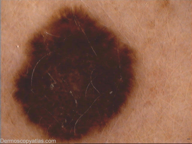
Description: Dermoscopy-Multiple brown dots with asymmetrical radial streaming.
History:
Presented for skin check.This mole(7 mm) on the left upper arm has been there for a long time without noticeable change in size or colour by the patient.Atypical looking naevus was excised due to dermoscopic findings of multiple brown dots and radial streaming.
Histopathology: Left deltoid- primary,non-ulcerated LEVEL 2 SUPERFICIAL SPREADING MELANOMA with regression arising in a dysplastic naevus.It shows active regression .The lesion is non-ulcerated and has Breslow thickness of approximately 0.7 mm.Dermal mitoses are present in small numbers indicating vertical growth phase.
This is an example of a spitzoid lesion . Rule out melanoma in lesions with asymmetrical spitzoid pattern.