
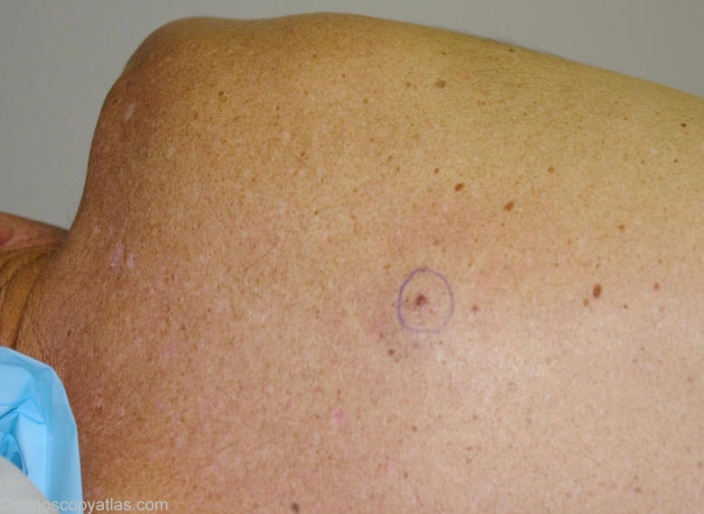
Site: Back
Diagnosis: Nevus dysplastic
Sex: M
Age: 57
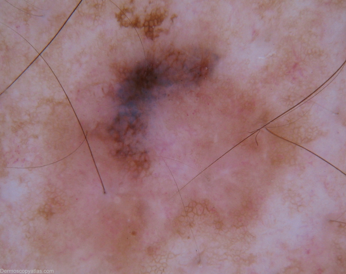
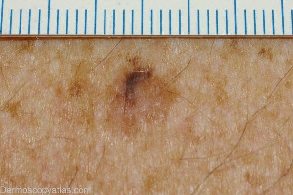
Type: Dermlite Non Polarised
Submitted By: Wynn Hlaing
Description: Clinical- Right infrascapular area,brown macule.
History:
This man presented for his skin examination .He was not aware of this pigmented lesion.He has no family or personal history of skin cancer.He has skin phototype II-III.
Regression structures may be found in melanoma and Clark naevus.In such instance the differentiation between these lesions may be difficult not only dermoscopically but also histopathologically .
Histopathology: Junctional dysplastic naevus exhibiting moderate dysplasia.Appearances fall short of in-situ melanoma.
This is an example of a lesion which has the same clinical appearance of all the other lesions from a distance,but at a closer look is different from the others.['Little Red Riding Hood' sign] IDS.