
Site: Calf
Diagnosis: Melanoma amelanotic
Sex: F
Age: 69
Type: Heine
Submitted By: Jean-Yves Gourhant
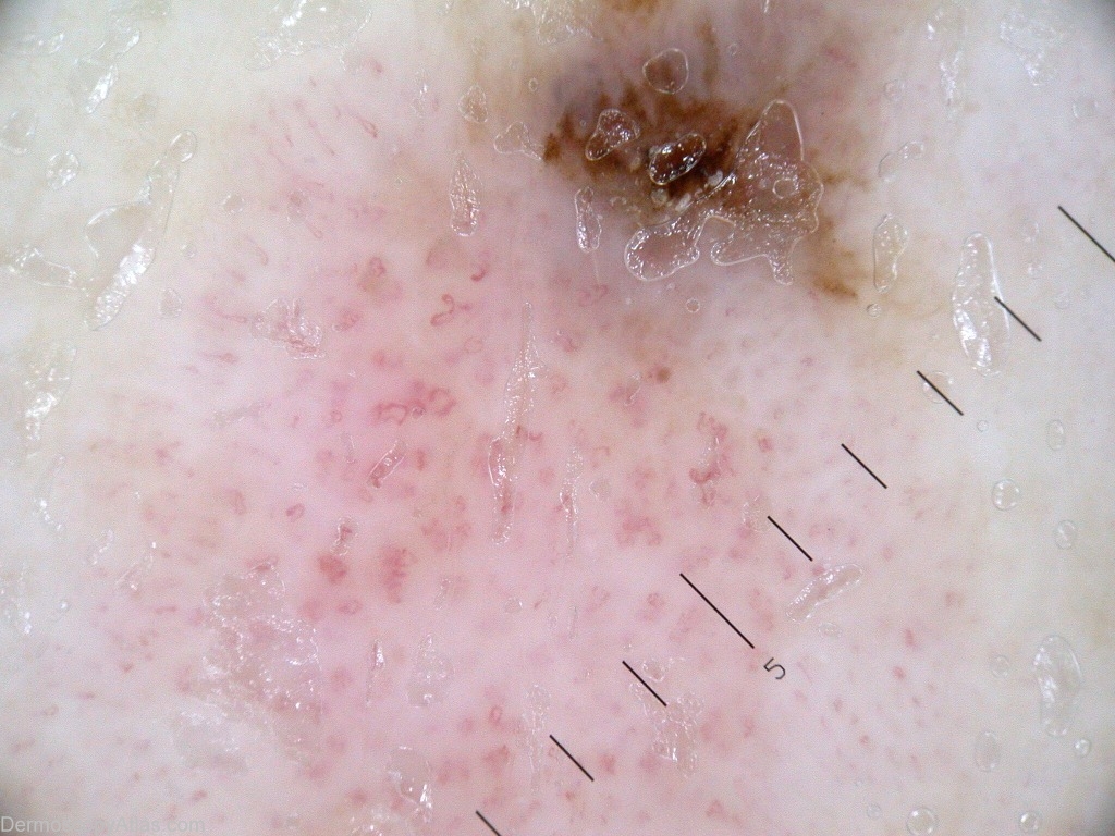
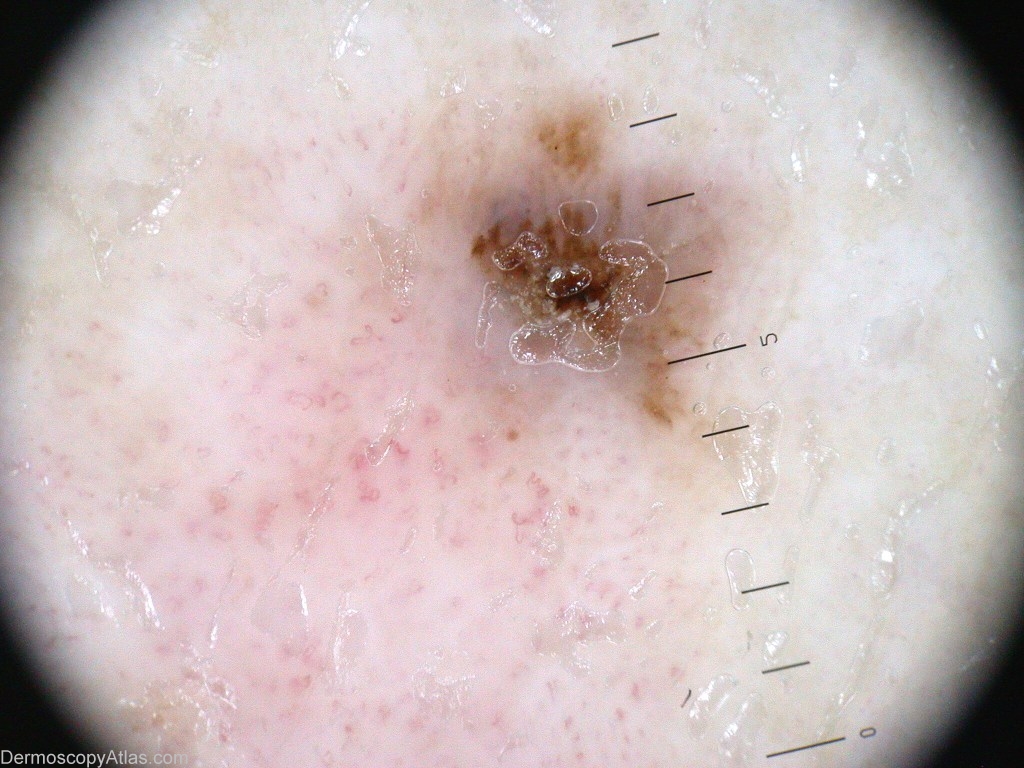
Description: The crusted area surrounded by grey-blue pigmentation. Below, the polymorphous vessels.
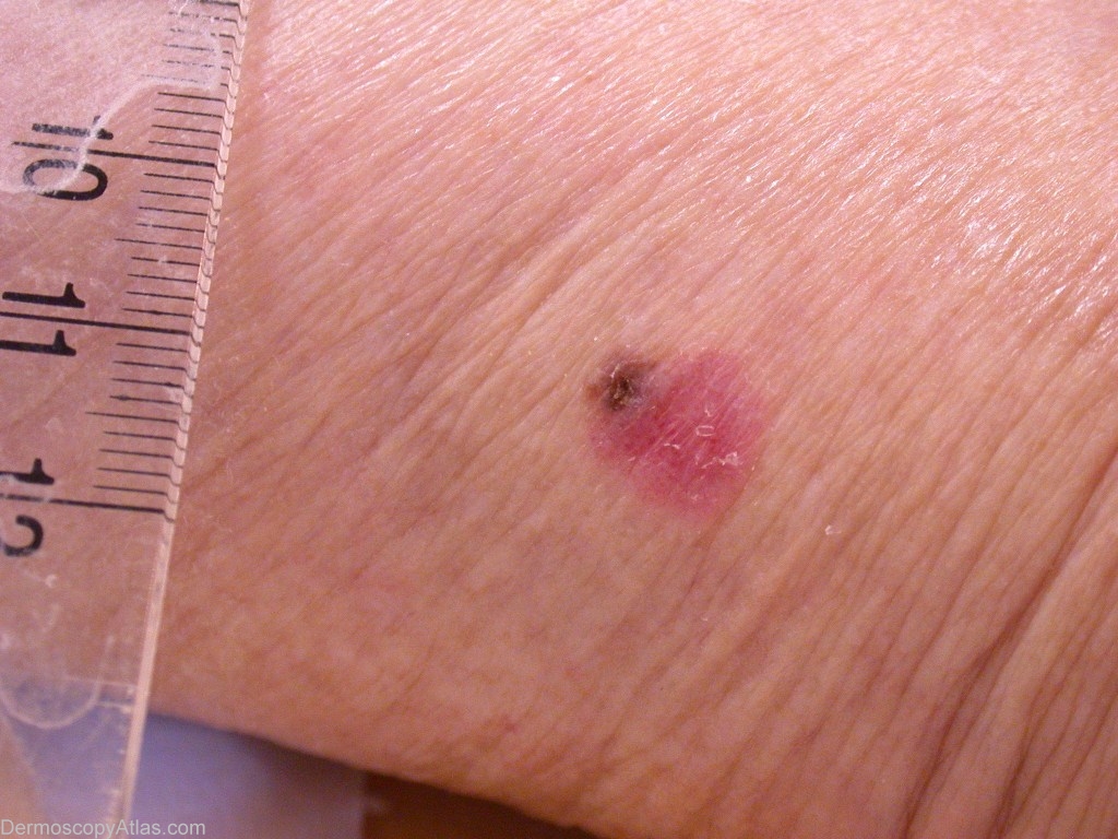
History: A 69 years old lady who came for this chronic lesion of the inferior part of the calf. No precise chronology. It was a pink, scaly, diameter 1 cm plaque, with a pigmented crust at his superior pole. Dermoscopy showed a polymorphous vascular pattern, with some corkscrew vessels, and for the pigmented pole, mainly a crust which was surrounded by blue-grey pigment.
The pathology revealed an invasive melanoma, SSM, Clark IV, Breslow 1.7.
Clue to diagnosis: Polymorphous vessels in association with areas of blue-grey pigment.