
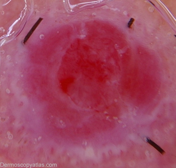
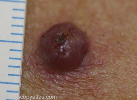
Site: Cheek
Diagnosis: Pyogenic granuloma
Sex: M
Age: 55
Type: Dermlite Non Polarised
Submitted By: Wynn Hlaing
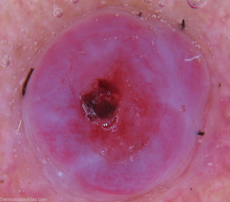
Description: Dermoscopy-White scaly collarette.
History:
This man presented with history of recurrent bleeding from his left cheek papule .It has been slowly enlarging for the past month.Second dermoscopic image was taken two weeks after the initial visit,before the curette and cauterization.Histopathology confirmed the clinical diagnosis of granuloma pyogenicum.
Important differential diagnosis for pyogenic granuloma is amelanotic melanoma.
A white scaly collarette likely corresponds to hyperplastic epithelium.