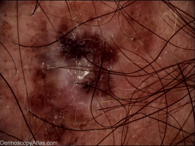
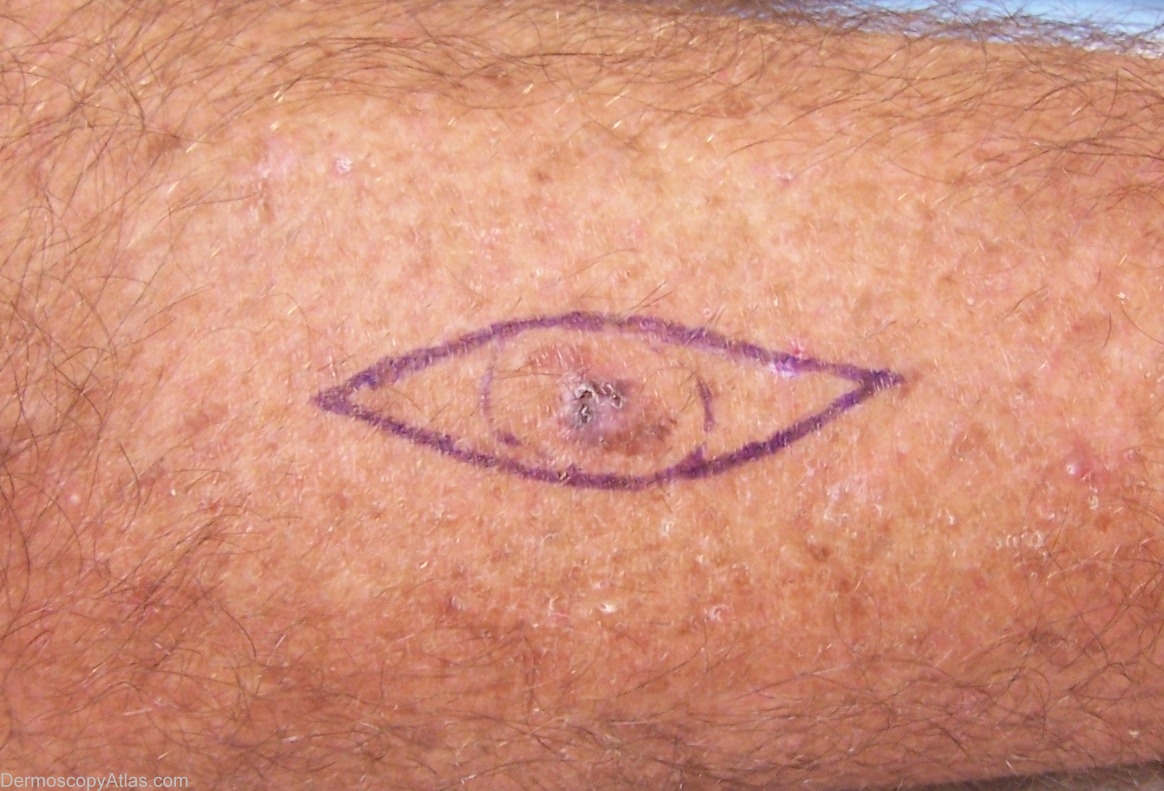
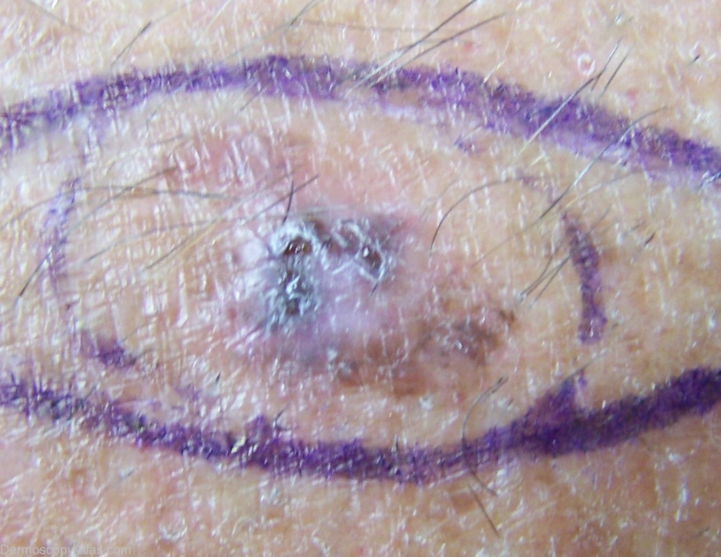
Loading
TeleDermConsult-logo
  • Recent Submissions
  • Search
  • Diagnosis
  • Quiz
  • Image comparison
  • Submit
  • Teaching Cases
  • Index
  • About

<< Back to Diagnosis Page

Image Number #2452 (Melanoma invasive)

Site: Leg

Diagnosis: Melanoma invasive

Sex: M

Age: 62

Type: Molemax

Submitted By: Greg Canning

View Full Size Image

Click Here to View Details

Description: Dermoscopy Mole Max , Streaks,

History: This 62 yr old builder has deeply tanned type 3 skin, and no history of skin cancer. He was not aware of this lesion on the posterior left calf. Excision showed SKIN POSTERIOR MEDIAL LEFT CALF - LEVEL 2, 0.70 MM THICK SUPERFICIAL SPREADING MALIGNANT MELANOMA.

DermNetNZ   eMedicine   PubMed   Dermatology Online   Archives   JAAD for "Melanoma invasive"

  • Recent Submissions
  • Search
  • Diagnosis
  • Quiz
  • Image comparison
  • Submit an Image
  • Teaching Cases
  • Index
  • About

Copyright 2022. Dermoscopy Atlas. ALL Rights Reserved.

Change Image