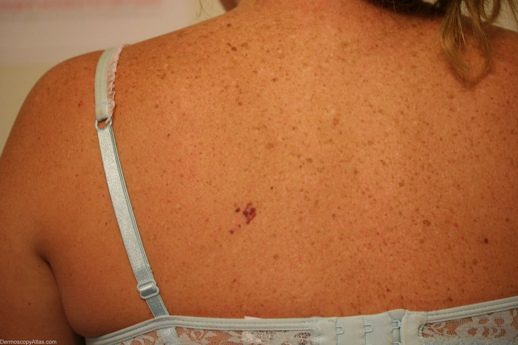
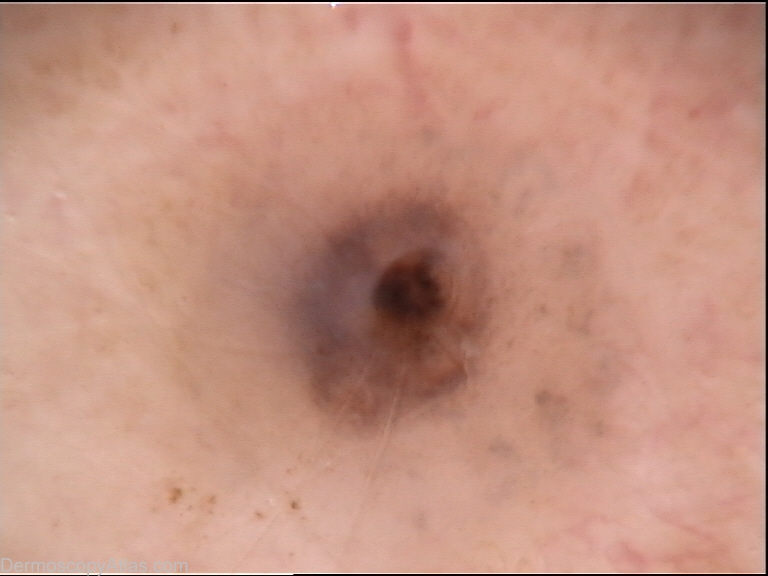
Loading
TeleDermConsult-logo
  • Recent Submissions
  • Search
  • Diagnosis
  • Quiz
  • Image comparison
  • Submit
  • Teaching Cases
  • Index
  • About

<< Back to Diagnosis Page

Image Number #2353 (Melanoma in situ)

Site: Back

Diagnosis: Melanoma in situ

Sex: F

Age: 28

Type: Mixed

Submitted By: Lester Cowell

View Full Size Image

Click Here to View Details

Description: Clincial

History:

Lesion present for over 12 months.  Family history of melanoma.

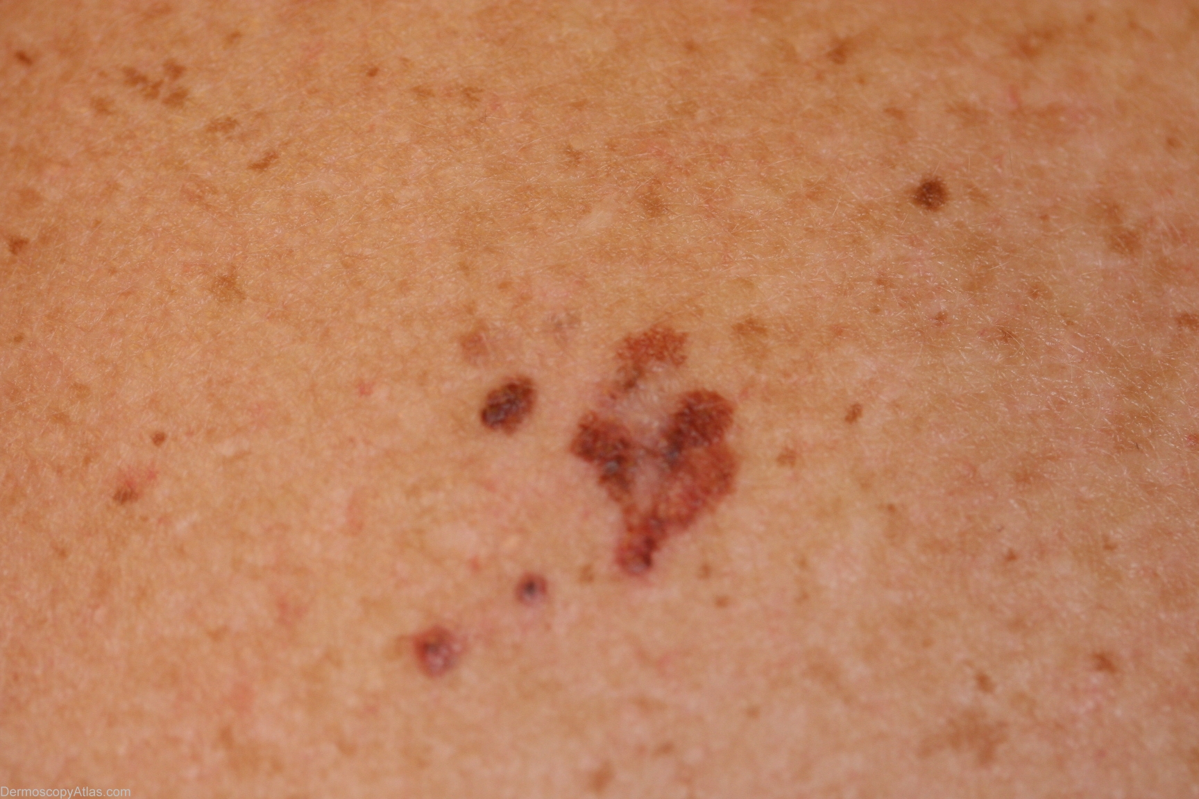
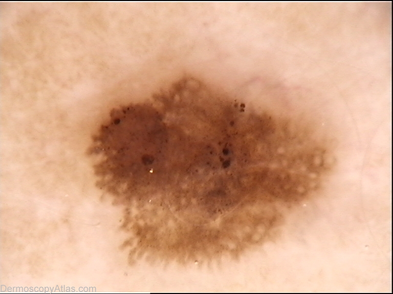
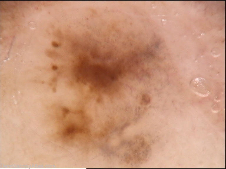
Close up by canon 350 D, ELMswith Fotofinder. at  x70.

Should this be described as an agminate melanoma ?

Lesion was excised and  smaller "satellites" were excised by encircling punch biopsy so each was assessed individually.

Each focus was reported as melanoma  - insitu x3  and invasive x1 . There were no features of in transit type lesions histologically in the "satellites". Each is a small melanoma with different structures.


DermNetNZ   eMedicine   PubMed   Dermatology Online   Archives   JAAD for "Melanoma in situ"

  • Recent Submissions
  • Search
  • Diagnosis
  • Quiz
  • Image comparison
  • Submit an Image
  • Teaching Cases
  • Index
  • About

Copyright 2022. Dermoscopy Atlas. ALL Rights Reserved.

Change Image