
Site: Leg
Diagnosis: Dysplastic nevus
Sex: M
Age: 56
Type: Heine
Submitted By: Lester Cowell
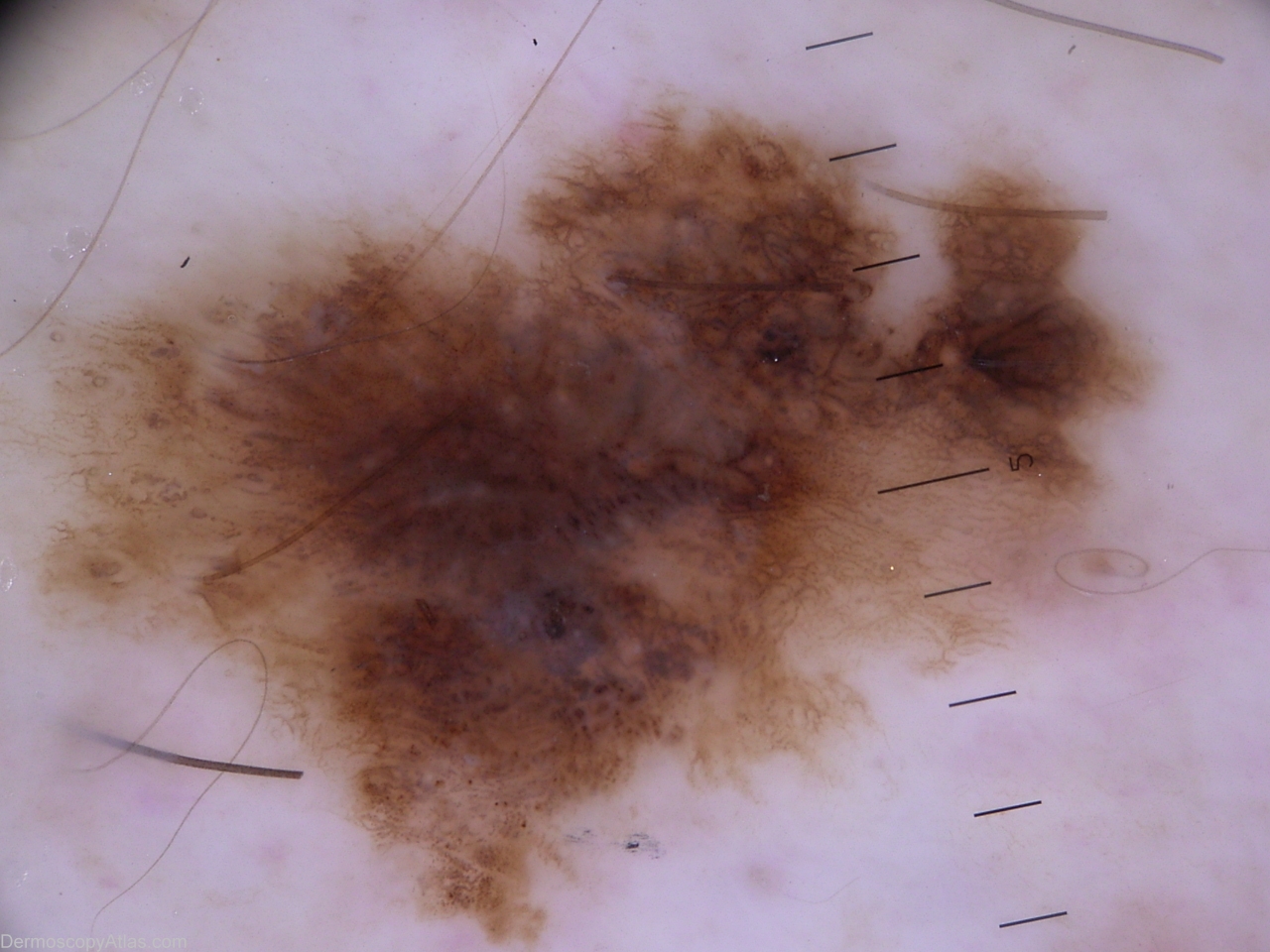
Description: lesion of distal 1/3 of medial leg border.
History:
Patient presented concerned with lesions that were clinically hyperkeratotic seborrheic keratoses.
Examination disclosed this solitary lesion that had been present many years but upon direct questioning had possibly changed in the last 6 - 12 months.
On this image, several reknowned dermoscopy authors at an international meeting were happy to call this melanoma insitu. Pathologists described this as dysplastic naevus with small foci of regression.
Dermoscopically it is an asymmetric combination of multiple patterns with multiple colors.
Structures include streaks, peripheral brown dots, globules, homogenous light brown areas, negative pigment network (6:00)