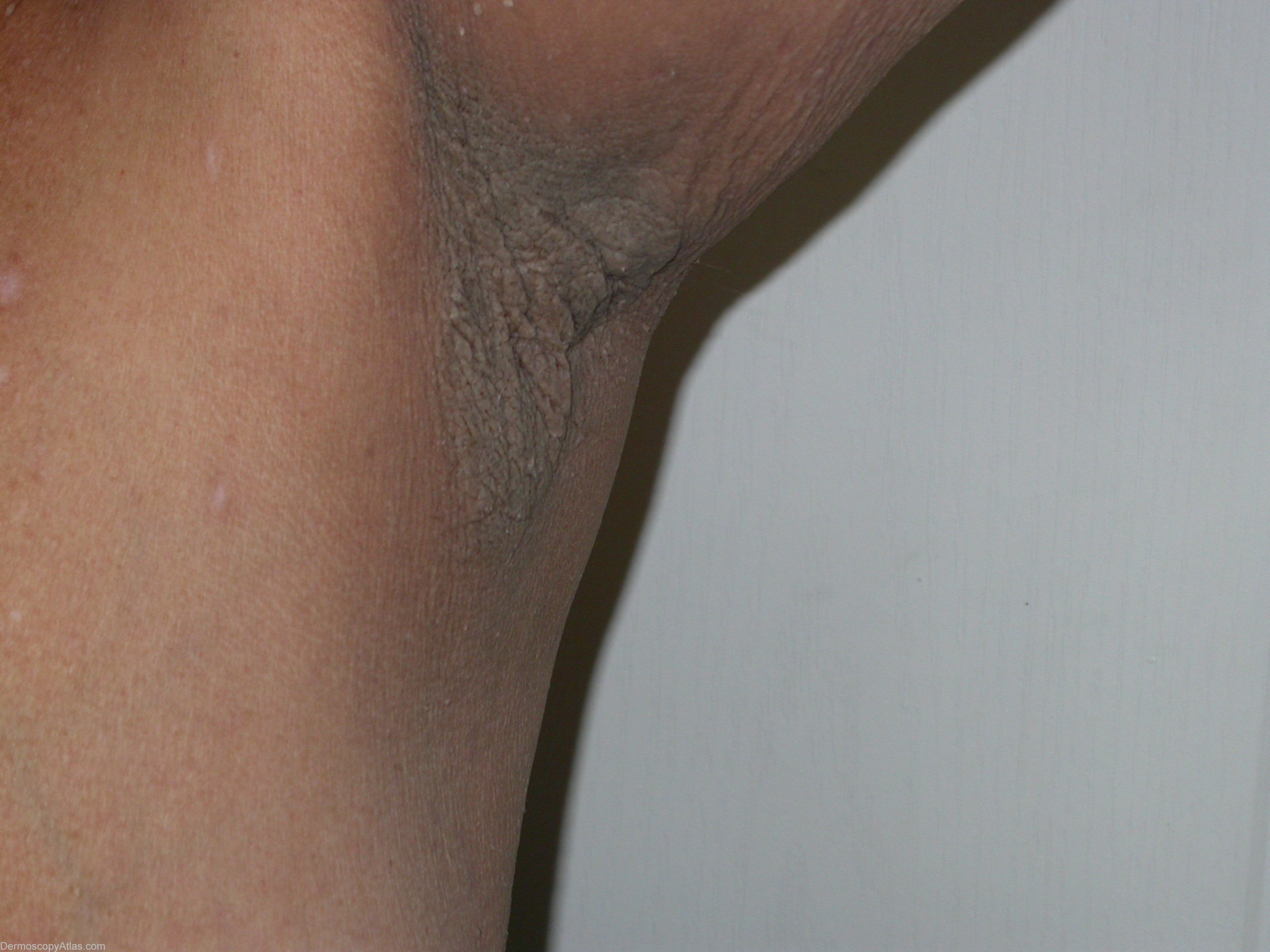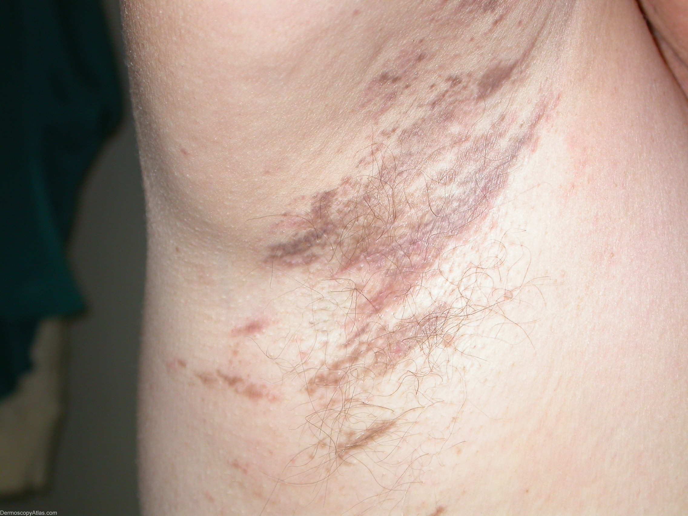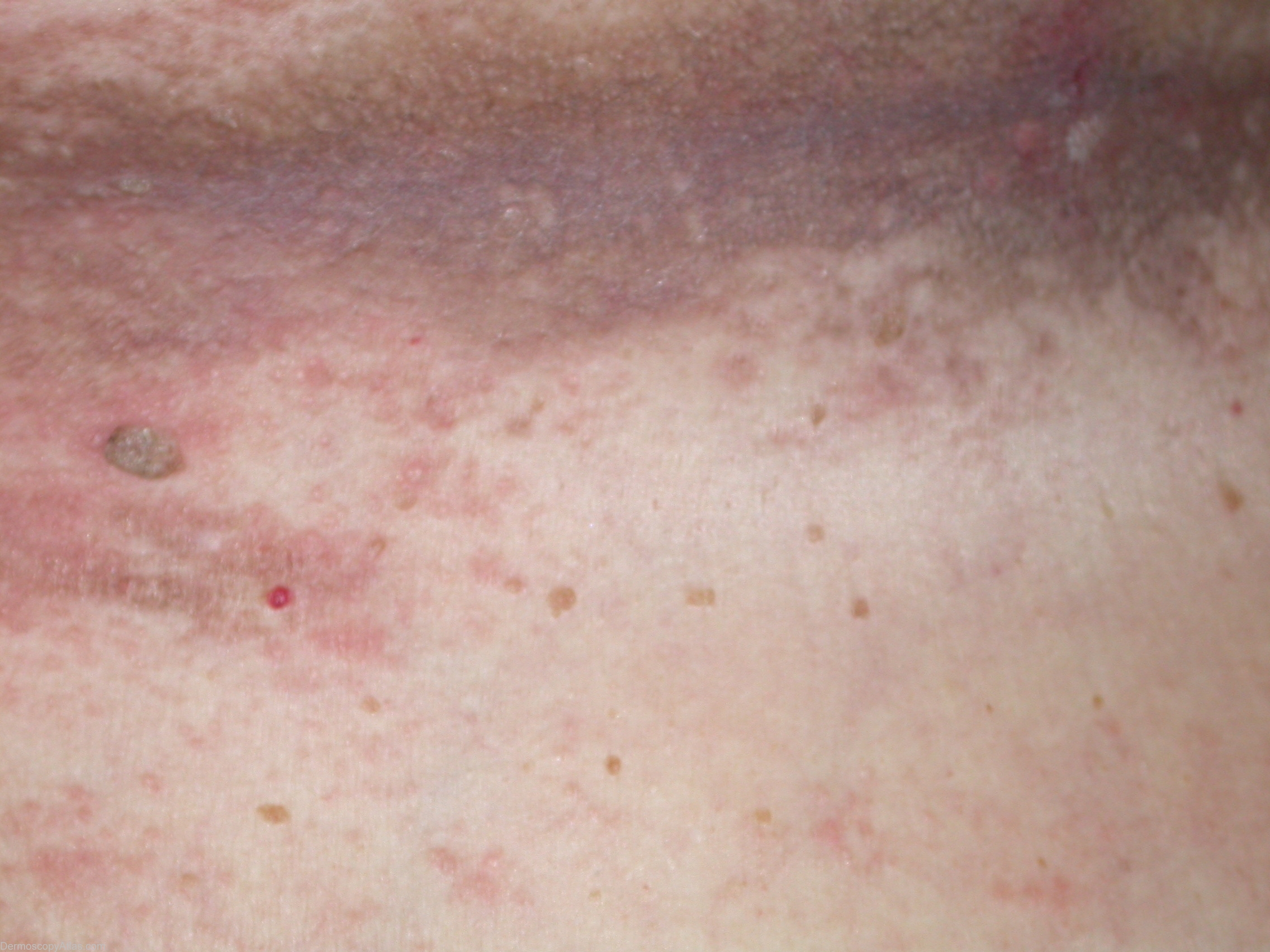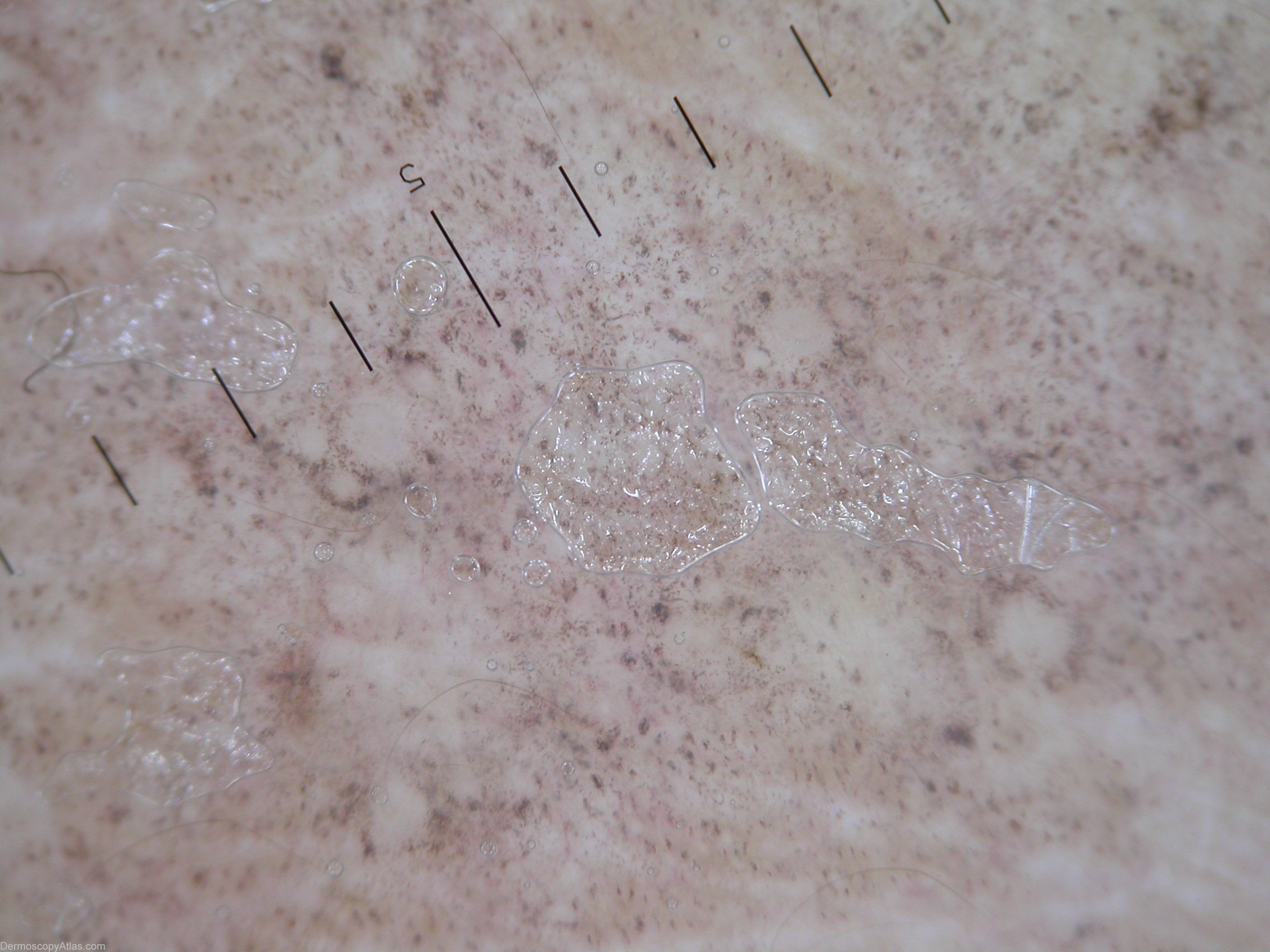
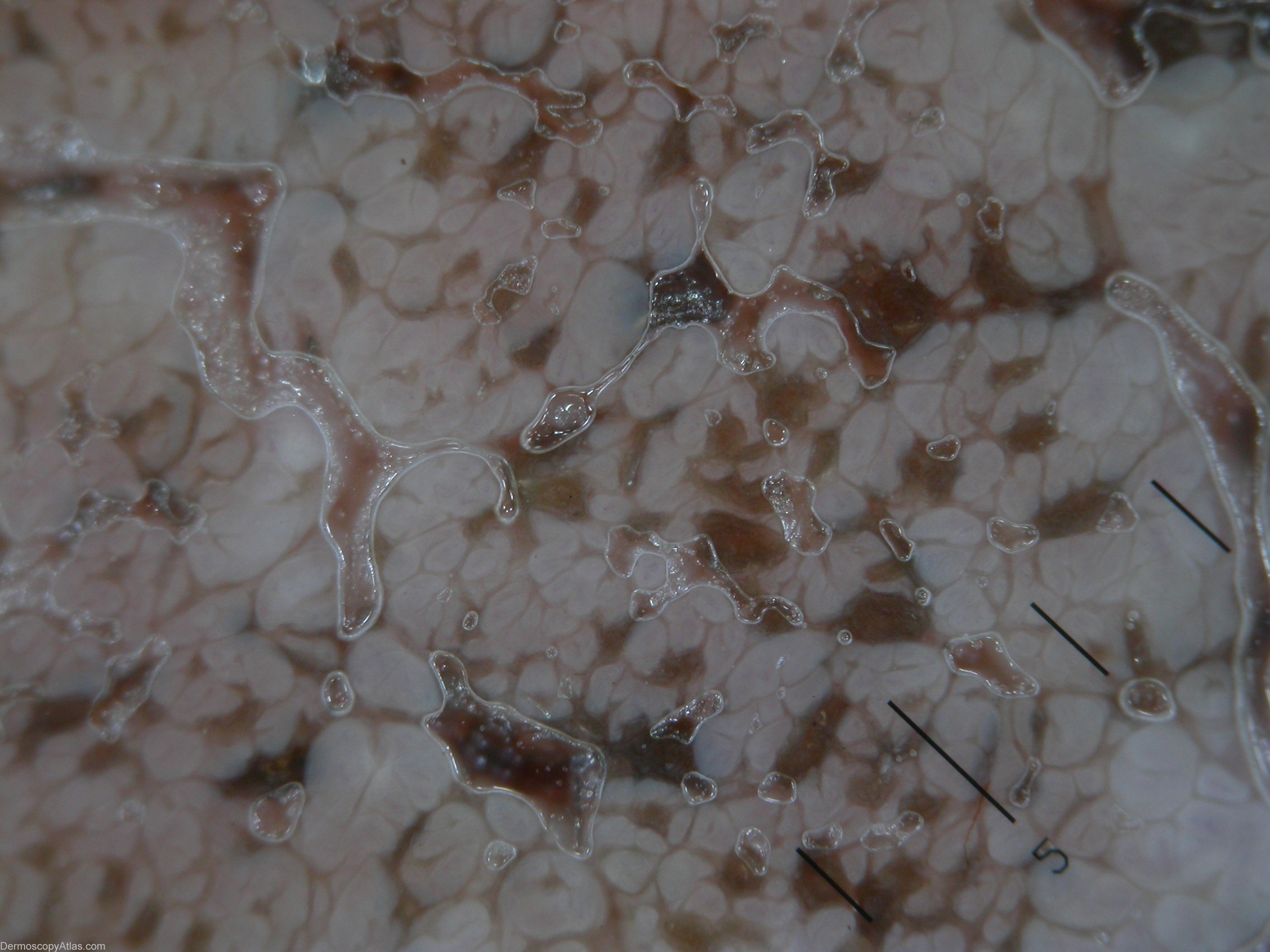
Site: Axillae
Diagnosis: Acanthosis nigricans v Lichen ruber planus
Sex: M
Age: 44
Type: Heine
Submitted By: Stelios Minas
Description: Exophytic papillary structures, multiple asymmetric blotches and milia like cysts.
History:
Differential diagnosis between Acanthosis nigricans(AN) and lichen ruber planus, pigmented form(LRP-PF) under dermoscopy.
This A 44-year-old woman developed persistent hyperpigmented velvety plaques in intertriginous areas of the axilla for a long time. Multiple brown skin tags are found in and around the affected areas.
She had high plasma insulin levels but a normal abdominal ultrasound.The skin lesions were treated with topical tretinoin 0.1% cream. AN
Dermoscopic description(AN): Exophytic papillary structures, multiple asymmetric blotches and milia like cysts.
This 57-old woman developed persistent hyperpigmented velvety plaques in intertriginous areas of the axilla 4 years ago. She was diagnosed as a Acanthosis nigricans. No other finding of an underlying malignancy or diabetes. Dermoscopic finding make us to think about LRP-PF.
Dermoscopic description(LRP-PF): Gray granules (pepper-like granules).
Our dermatoscopic findings allowed us to make the correct diagnosis of AN and make it easier to differentiate it from lichen ruber planus, pigmented form.
As you see from these 2 cases clinical images of AN and lichen ruber planus, pigmented form are very similar and dermoscopy can make the differentiation easier.
Differential diagnosis between Acanthosis nigricans and lichen ruber planus, pigmented form under dermoscopy.: For AN dermoscopic description is Exophytic papillary structures, multiple asymmetric blotches and milia like cysts and oppositive site for LRP-PF is gray granules (pepper-like granules) and absence of dermoscopic criteria for AN.
