
Site: Abdomen
Diagnosis: Nevus recurrent
Sex: F
Age: 39
Type: Heine
Submitted By: Stelios Minas
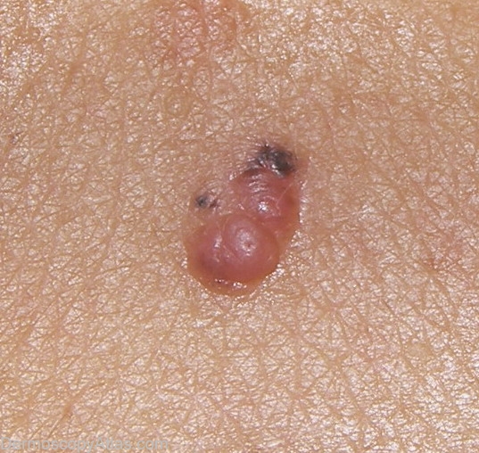
Description: Clinical of lesion at site of previous laser removal of a "nevus"
History:
The patient has a history of an excised pigmented lesion done with laser a year ago. Some time after this she has developed a new lesion precisely at the excision site. No previous histopathology report (laser excision). I have seen very often recurrent nevus after laser excision. Laser excision is not recommended for melanocytic lesions.
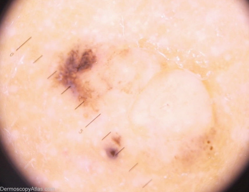
As Ian McColl said:Always check the original histopathology of a previous excision. In benign recurrences the pigment usually does not extend beyond the scar margins. See this reference
Also this Google Book Reference gives access to an online histopathology reference by Phil Le Boit