
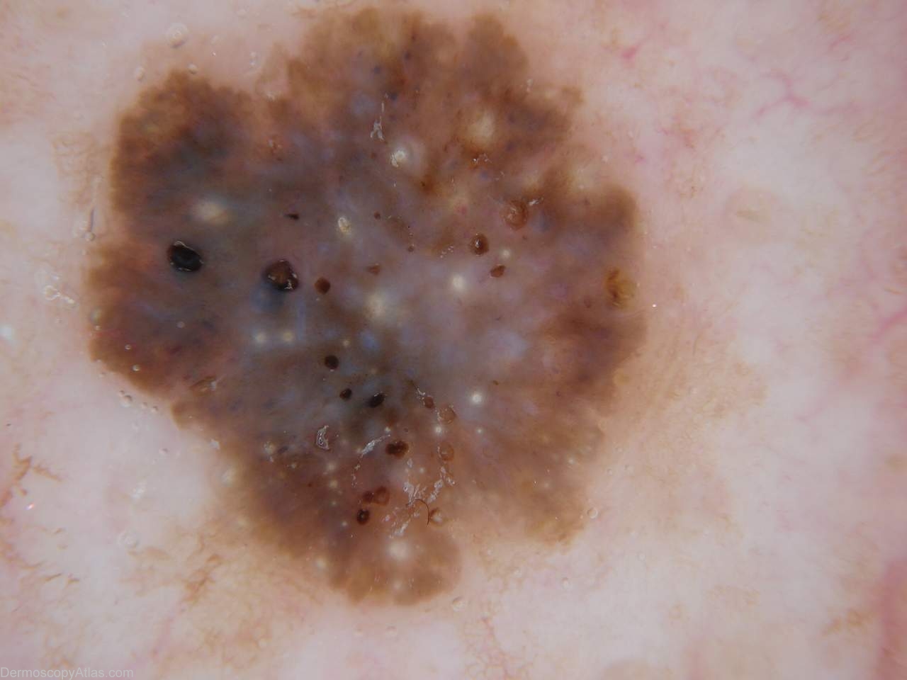
Site: Back
Diagnosis: Seborrhoeic keratosis
Sex: M
Age: 67
Type: Dermlite Non Polarised
Submitted By: Ian McColl
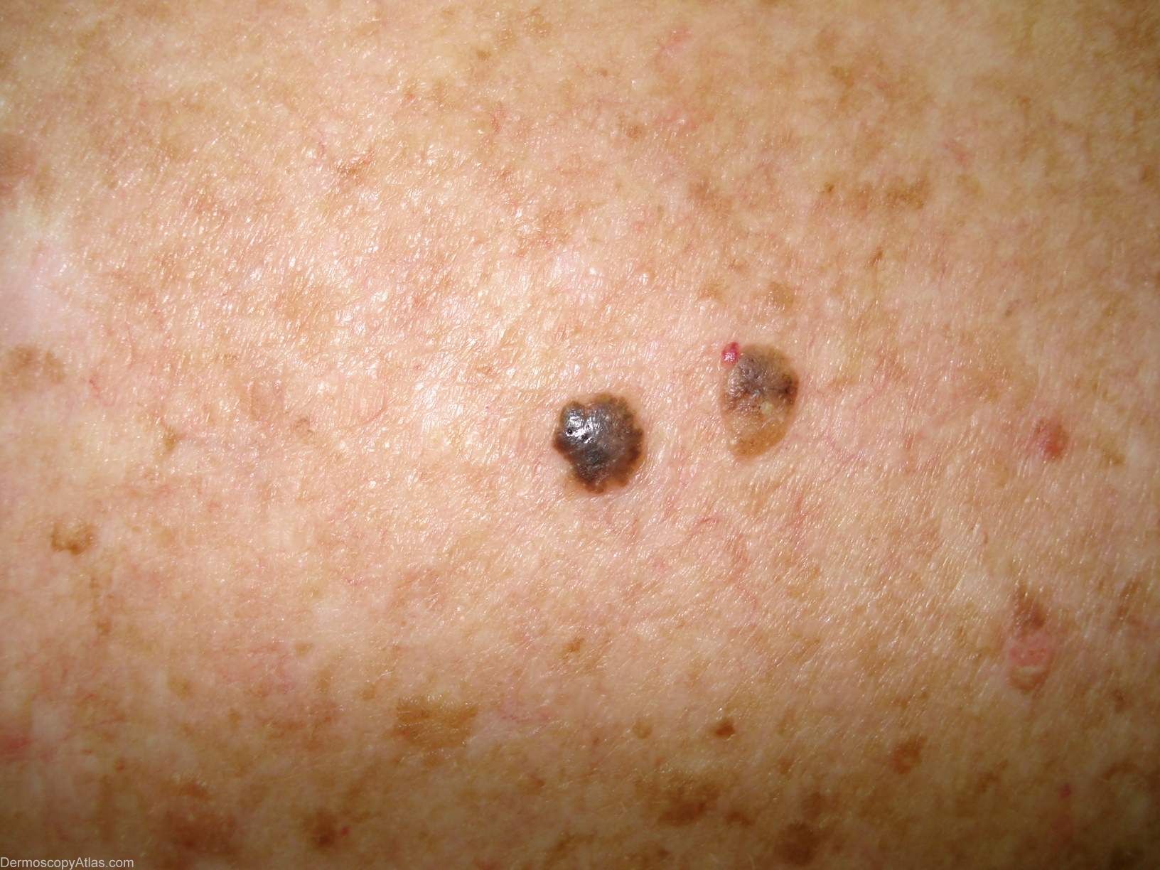
Description: Dermoscopy showing milial cysts and comedo openings. There are also homogeneous pigmented areas.
History:
This seborrhoeic keratosis shows many of the classical features of this type of lesion. The borders are usually sharp with numerous commedo like openings, crypts and fissures. Pigmented areas are often stuctureless. In Harald Kittler's terminology there are many clods of varying colours with orange predominating. Milia like cysts are another common feature of seborrhoeic keratoses. Occassionaly a less pigmented area may show underlying hairpin like vessels.