
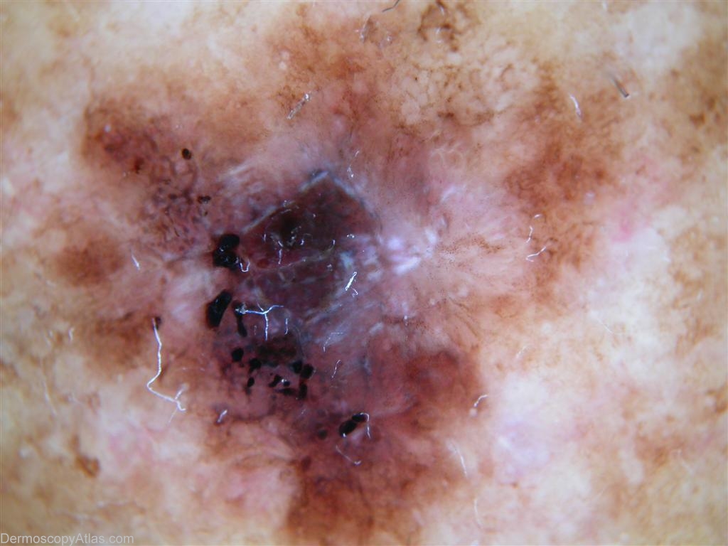
Site: Leg
Diagnosis: Melanoma in situ
Sex: F
Age: 68
Type: Heine
Submitted By: Keith Hopkins
Description: Dermoscopic view showing a surrounding pigment network with multiple black dots and structureless black pigment. There is also a blue grey veil.
History:
Fitzpatrick type 3 skin, sun worshipper. Lesion noted to be enlarging over three months.
Dermoscopic examination
3 point check list 3/3 (Asymmetry, Atypical network and Blue-white structures).
Pattern Analysis
Global-Non specific pattern and a multi-component appearance (areas of network, dots and globules, and diffuse hyperpigmented and hypopigmented areas).
Melanoma specific local features include atypical network, irregular dots and globules, irregular blotches and blue-white structures.
Therefore both 3 point checklist and pattern analysis suggestive of melanoma.
Other dermoscopic features
5 or more colours are present.
White scar liked regressed areas. Homogeneous purple and black areas.