
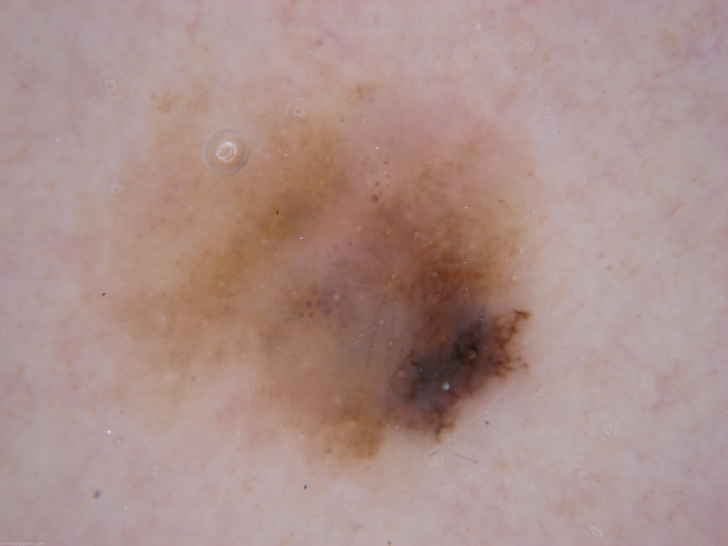
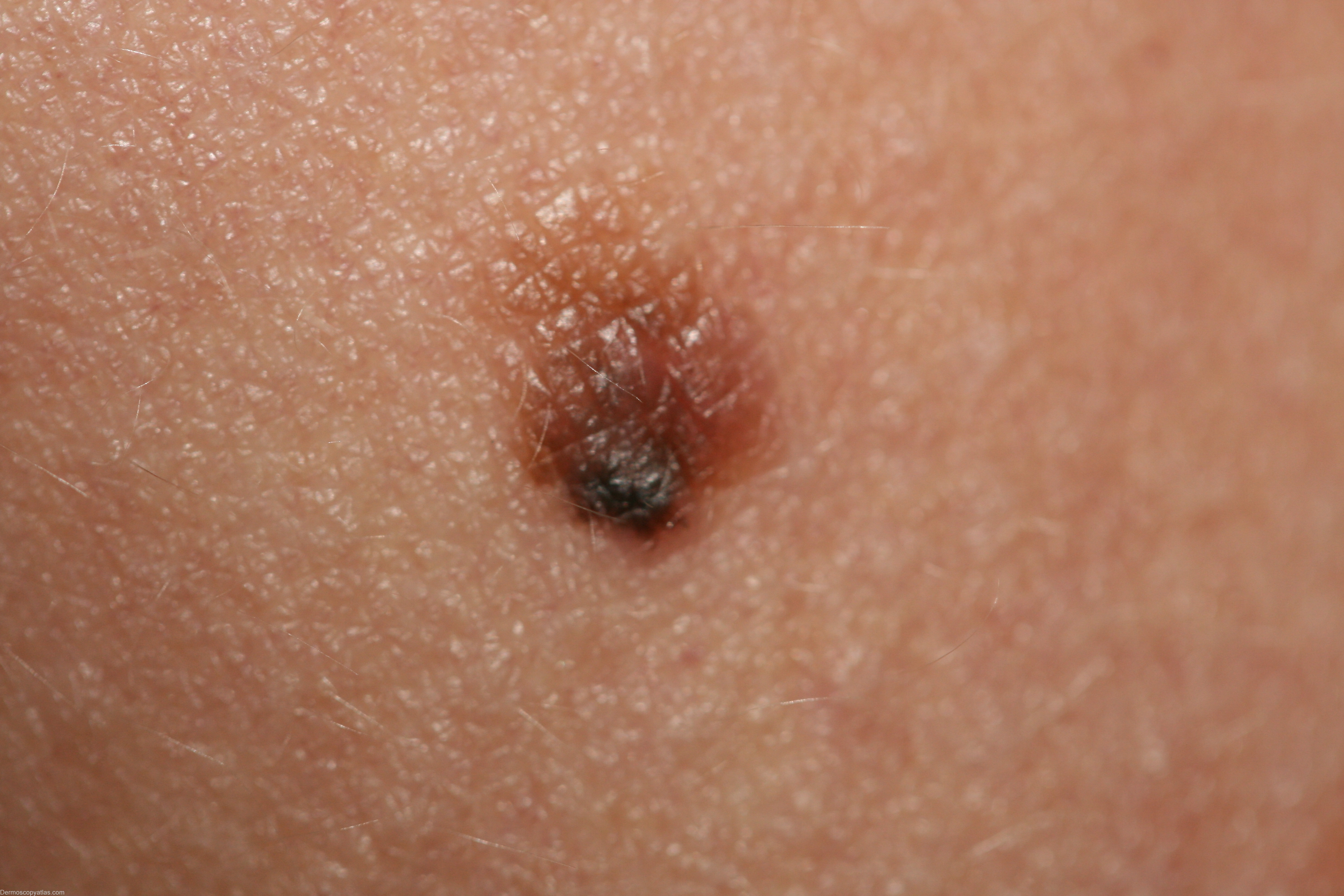
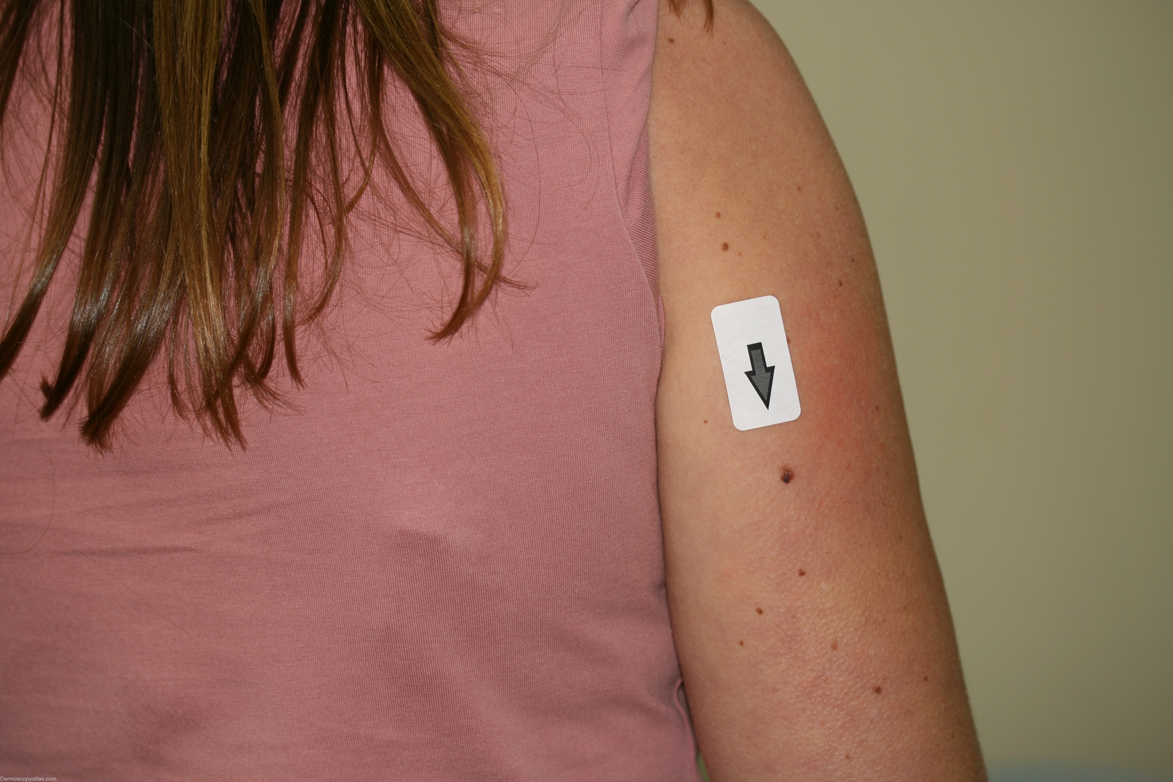
Site: Arm,upper
Diagnosis: Melanoma invasive
Sex: F
Age: 42
Type: Dermlite Non Polarised
Submitted By: Cliff Rosendahl
Description: This melanoma has inverse network and streaks which are borderline for meeting the criteria of pseudopods. There are also areas of blue-grey colour. The melanoma is quite asymmetrical and has several colours.
History: This 42 year old lady presented to obtain a referral to a gastroenterologist. This lesion was noted on the back of her arm as she walked into the consulting room. It was removed at that time as an excision biopsy with 2mm margins and histology confirmed a level 2 superficial spreading melanoma 0.4mm Breslow thickness.