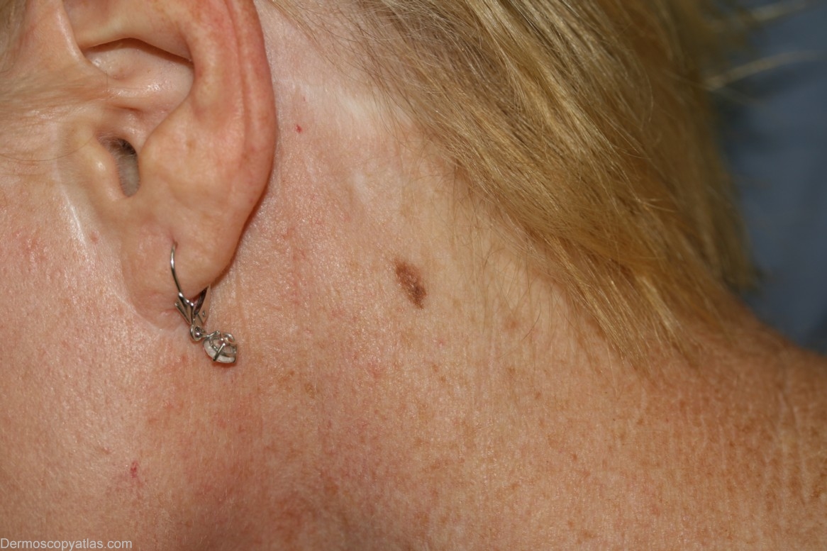
Loading
TeleDermConsult-logo
  • Recent Submissions
  • Search
  • Diagnosis
  • Quiz
  • Image comparison
  • Submit
  • Teaching Cases
  • Index
  • About

<< Back to Diagnosis Page

Image Number #2287 (Melanoma in situ)

Site: Neck side

Diagnosis: Melanoma in situ

Sex: F

Age: 58

Type: Dermlite Polarised

Submitted By: Cliff Rosendahl

View Full Size Image

Click Here to View Details

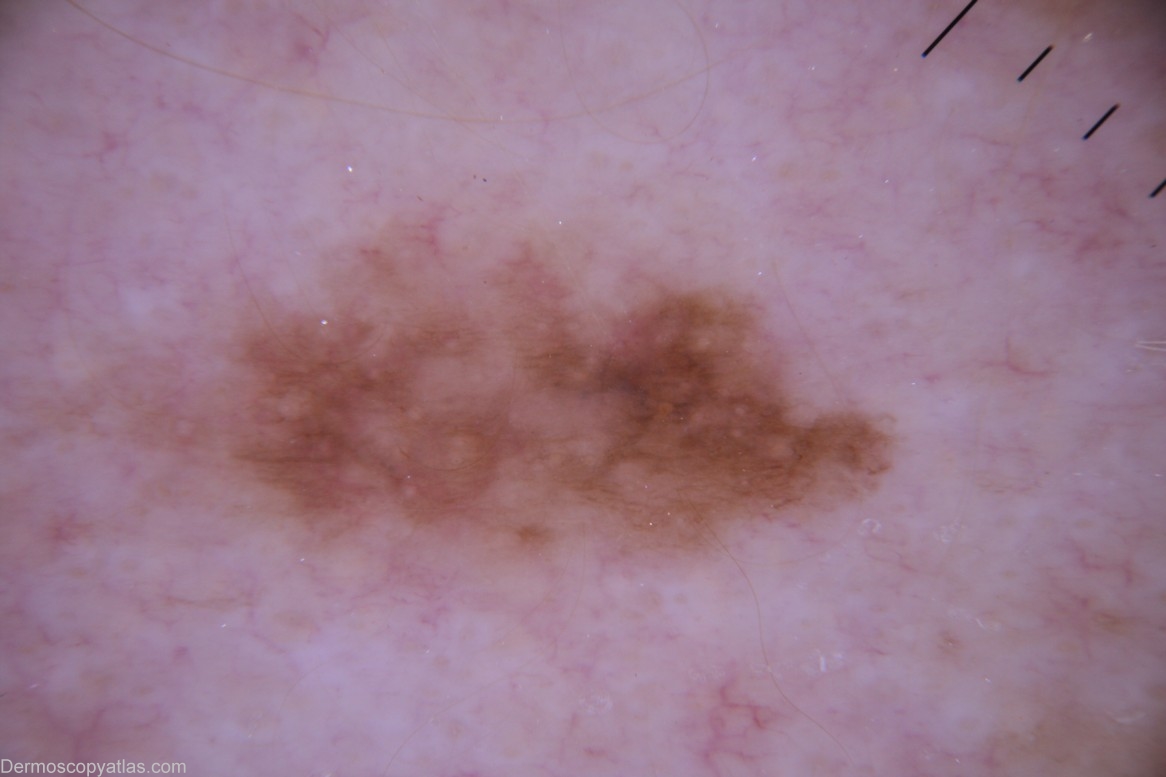
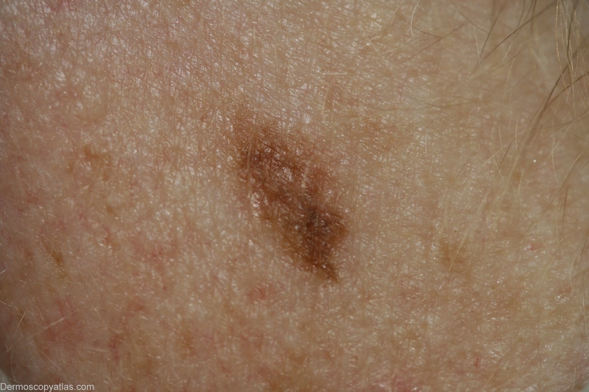
Description: Abrupt border

History: This 58 year old lady with no past history of skin malignancy was having a general medical examination. She was not concerned by this lesion but when interrogated she thought it had only appeared in the "last few years". I thought that the pigment ended abruptly at the inferior border and dermoscopically there was a hint of blue-grey just superior to this. The pathologist confirmed that the "melanoma" histology was confined to that focus.

DermNetNZ   eMedicine   PubMed   Dermatology Online   Archives   JAAD for "Melanoma in situ"

  • Recent Submissions
  • Search
  • Diagnosis
  • Quiz
  • Image comparison
  • Submit an Image
  • Teaching Cases
  • Index
  • About

Copyright 2022. Dermoscopy Atlas. ALL Rights Reserved.

Change Image