
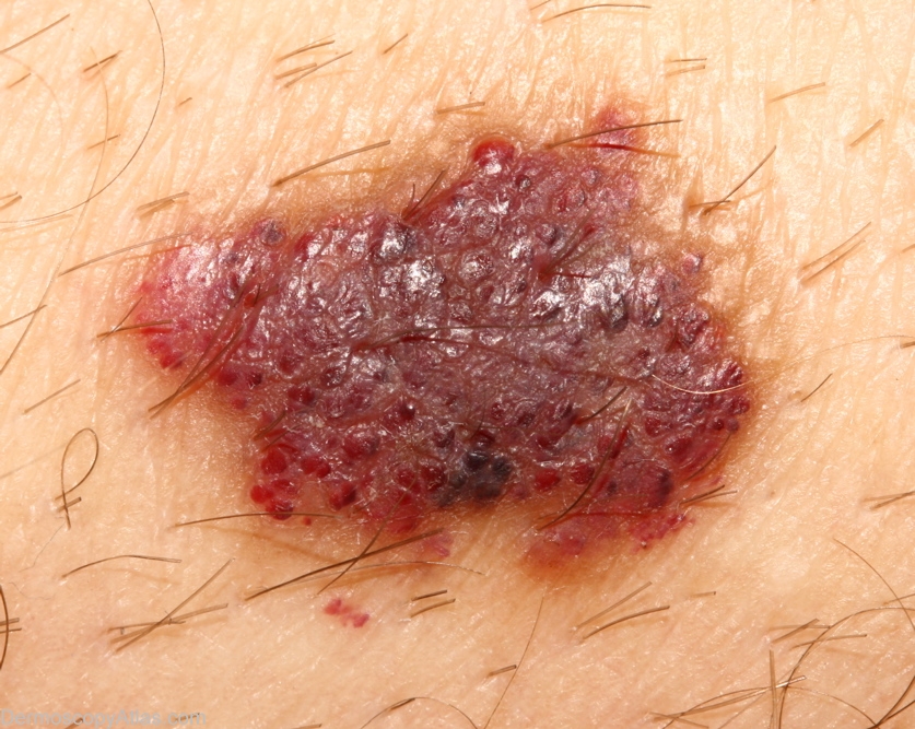
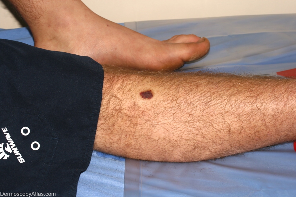
Site: Shins
Diagnosis: Haemangioma
Sex: M
Age: 24
Type: Dermlite Non Polarised
Submitted By: Alan Cameron
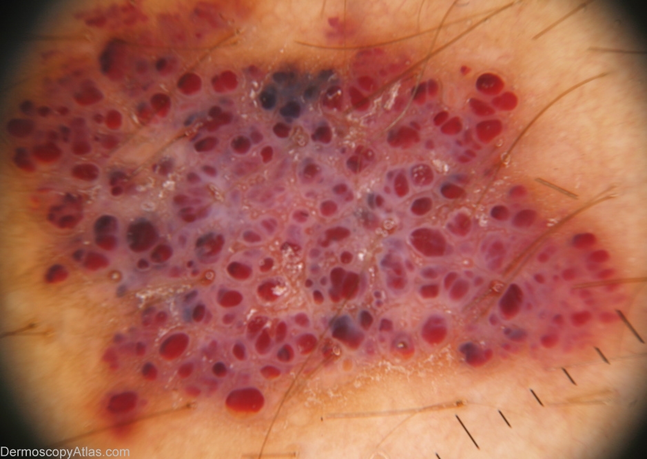
Description: macro. Note the dark area which might raise the suspicion of melanoma. This area will appear red/ blue under the dermatoscope.
History:
Lesion on the right shin. Some haemangiomas such as this one can have dark areas within them that may suggest melanoma. These vascular areas are however red/ blue under dermatoscopy. Some haemangiomas may have white scar like areas separating the vascular spaces. When a haemangioma is involuting there may be a prominent central white scar.
Important recent article on the pathogenesis of infantile haemangiomas