
Site: Calf
Diagnosis: Melanoma regression
Sex: F
Age: 72
Type: Heine
Submitted By: Ian McColl
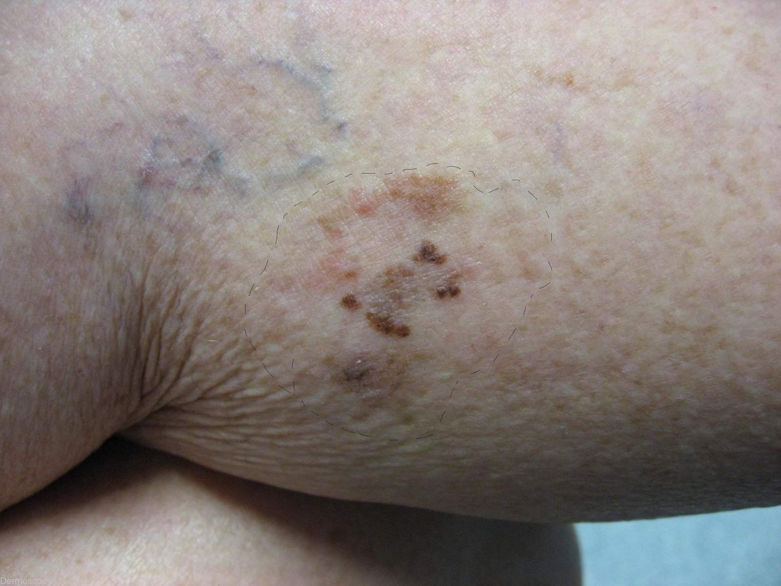
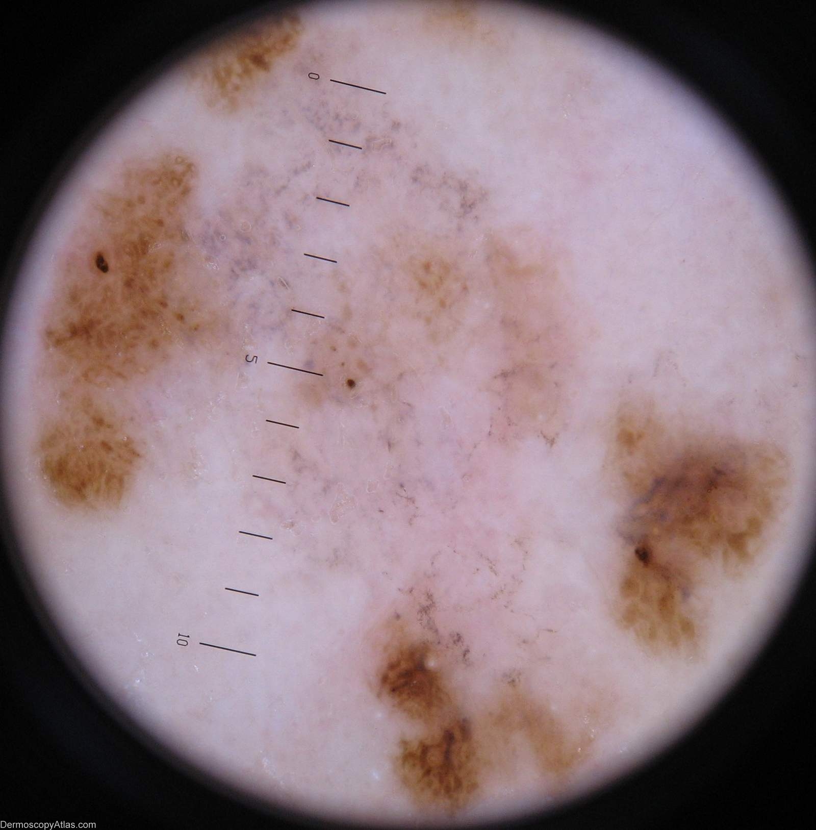
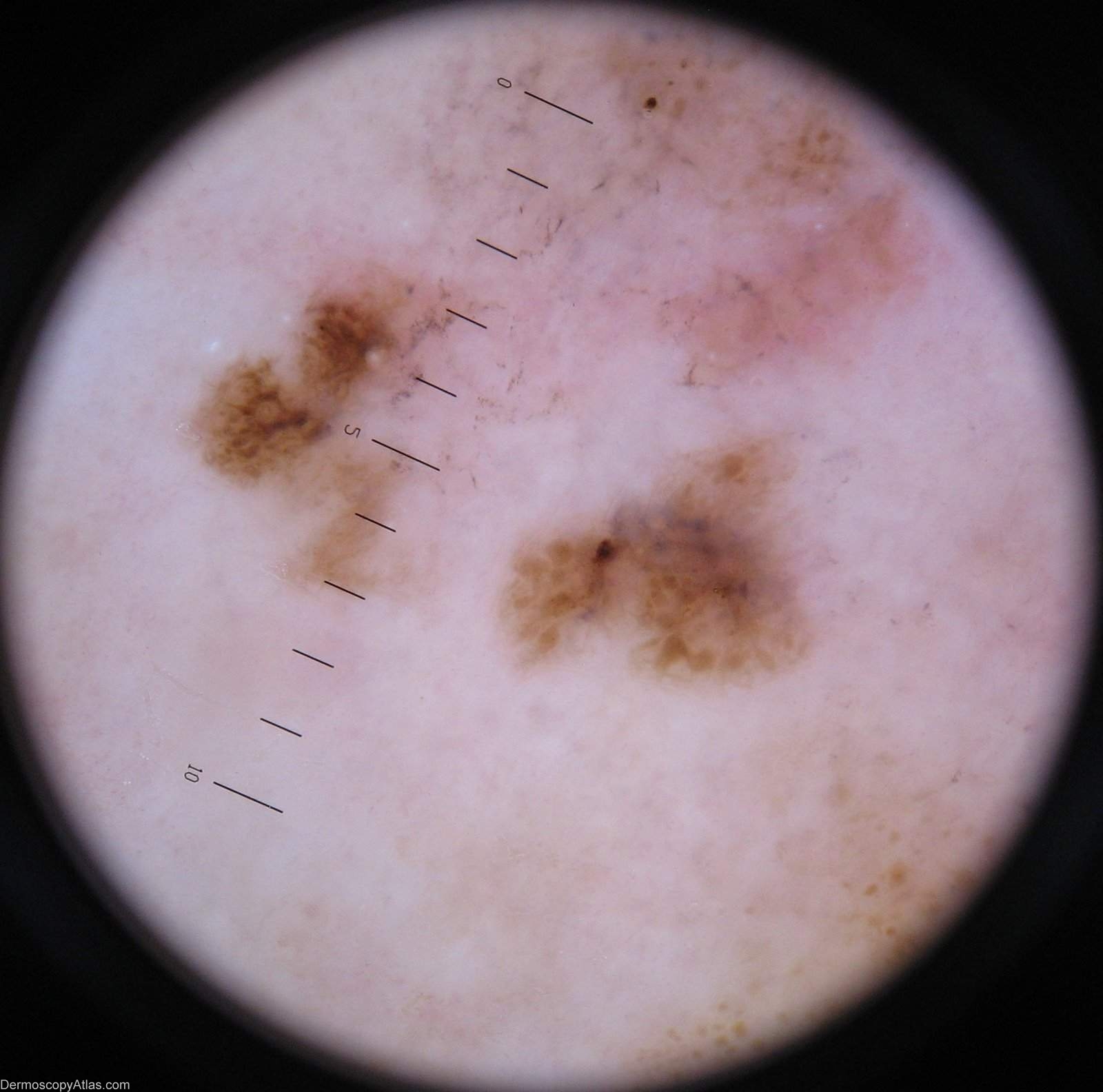
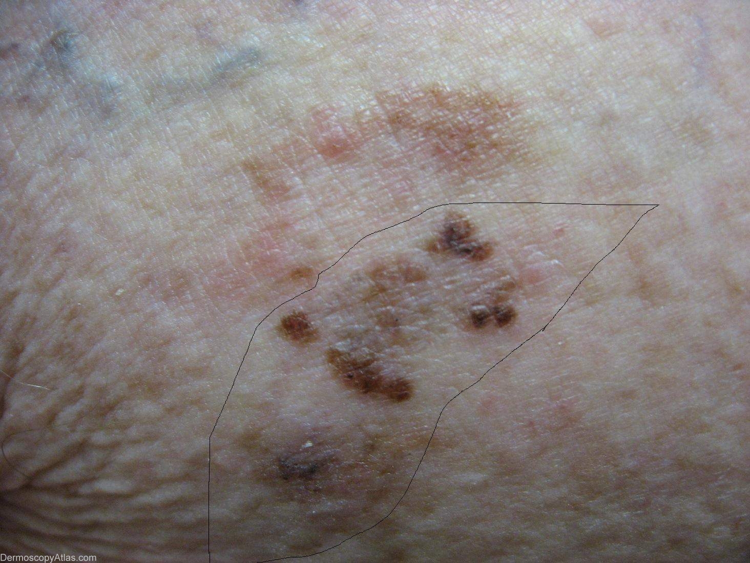
Description: Area for incisional biopsy. This shows the grey area of regression between the residual pigmented lesion.
History:
Case courtesy of Dr Greg Canning This poorly demarcated lesion is approx 5 cm in diameter and has resided on the left proximal postero medial calf of it's 72 year old owner for as long as she can remember. On reflection however she thinks it may have enlarged somewhat over the years. She has been repeatedly reassured about it by the doctors who have treated her numerous non melanoma skincancers.
Histopathology of incisional biopsy There is an atypical melanocytic lesion composed of disparate foci separated by zones of regression. There are moderately to markedly atypical melanocytes forming irregular junctional nests and invading the epidermis. A lichenoid dermal inflammatory infiltrate is present and there are two foci of superficial nvasion of the dermis to the interface of papillary and reticular dermis. The Breslow thickness in each case is 0.4 mm. Mitoses are rare. The minimum clearance of the invasive components is 2.5 mm from the 3 o'clock margin. Atypical junctional nests almost abut the 3 o'clock edge.
The degree of regression obfuscates interpretation of the precursor lesion, but lentiginous activity is seen and involvement of a hair follicle. In combination with the multifocal nature of the lesion it
is likely that this is lentigo maligna melanoma.
View the Bog discussion of this case.