
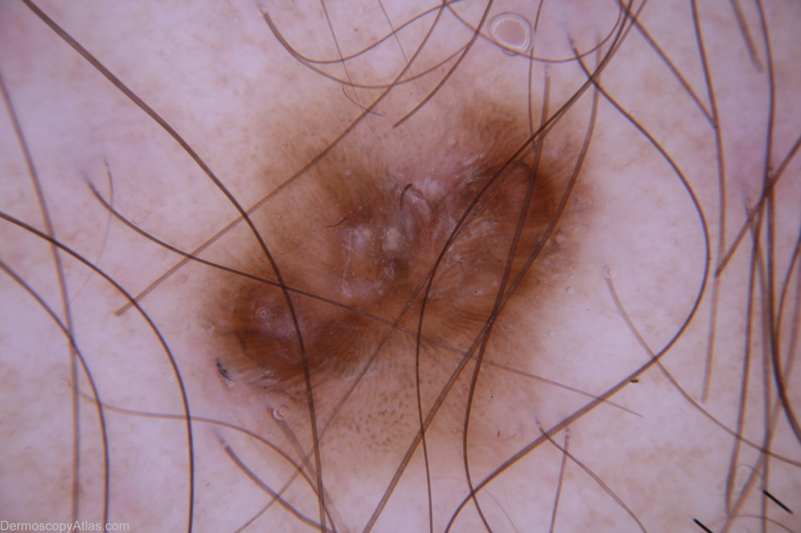
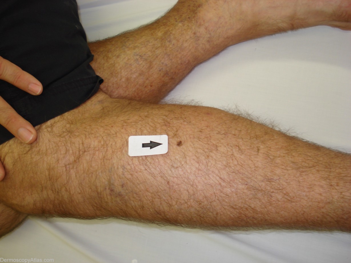
Site: Leg
Diagnosis: Large cell acanthoma
Sex: M
Age: 54
Type: Dermlite Non Polarised
Submitted By: Cliff Rosendahl
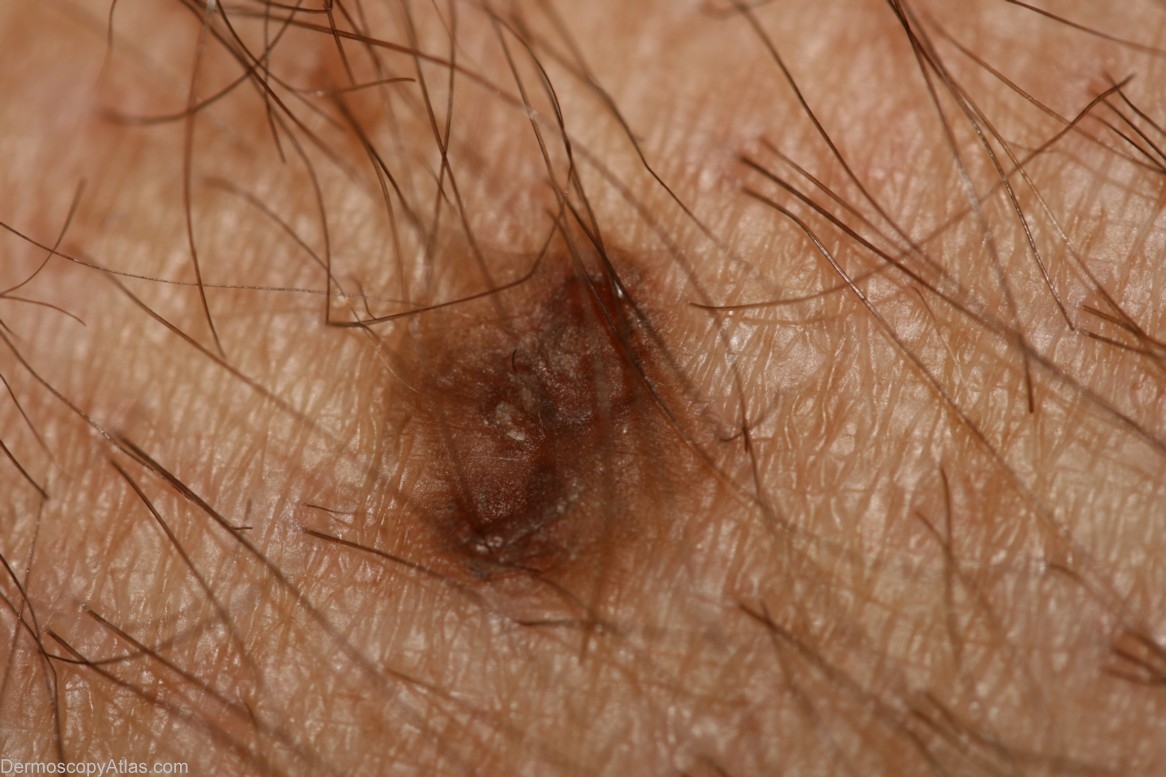
Description: Dermoscopy
History: This man presented for a regular skin check. This lesion although believed to be benign could not be confidently diagnosed so was subjected to shave-removal biopsy.
"Large cell acanthoma occurs as a sharply demarcated,often lightly pigmented patch,approximately 3-10 mm in diameter on the sun-exposed skin of middle-aged and elderly individuals. It is usually solirtary. Clinically it resembles a seborrhoeic or actinic keratosis. Large cell acanthoma is thought to comprise sunlight induced clones of abnormal cells without a tendency to malignancy. As such it is a distinctive condition and not a variant of solar lentigo..."(Skin Pathology,David Weedon)
View the Blog discussion of this case