
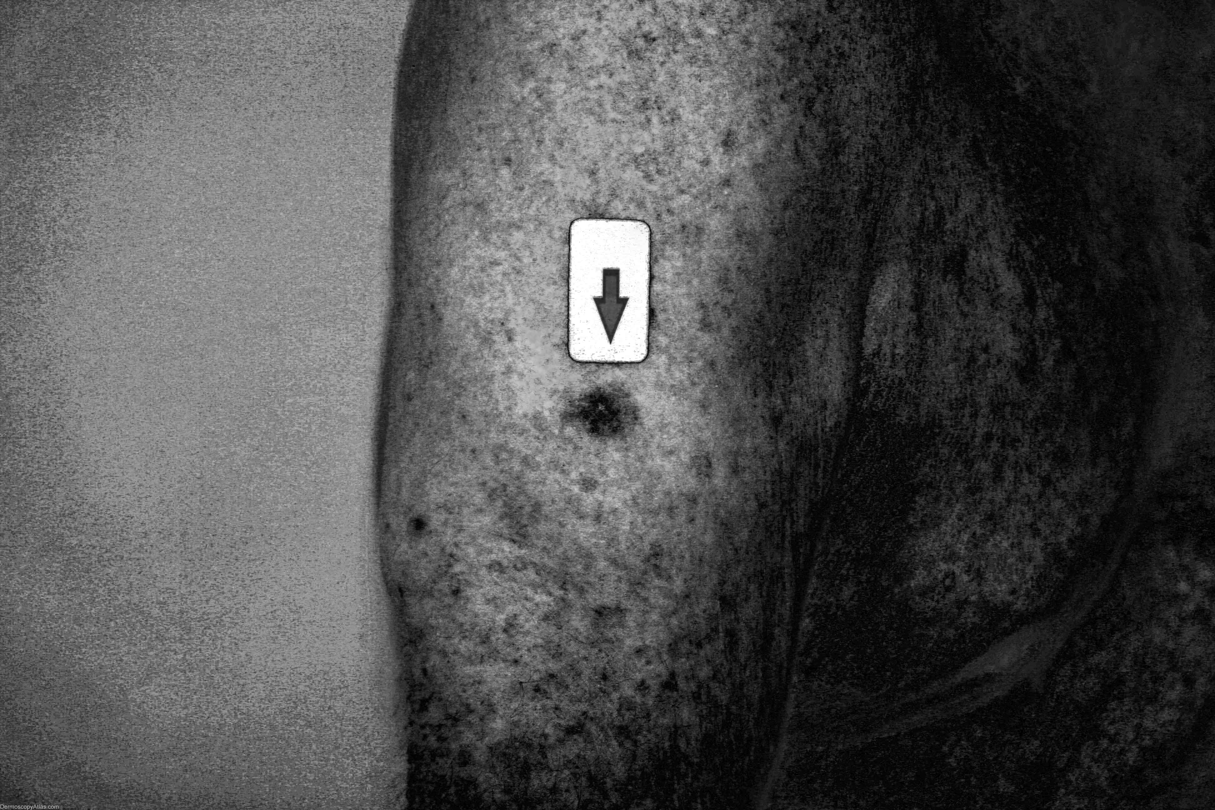
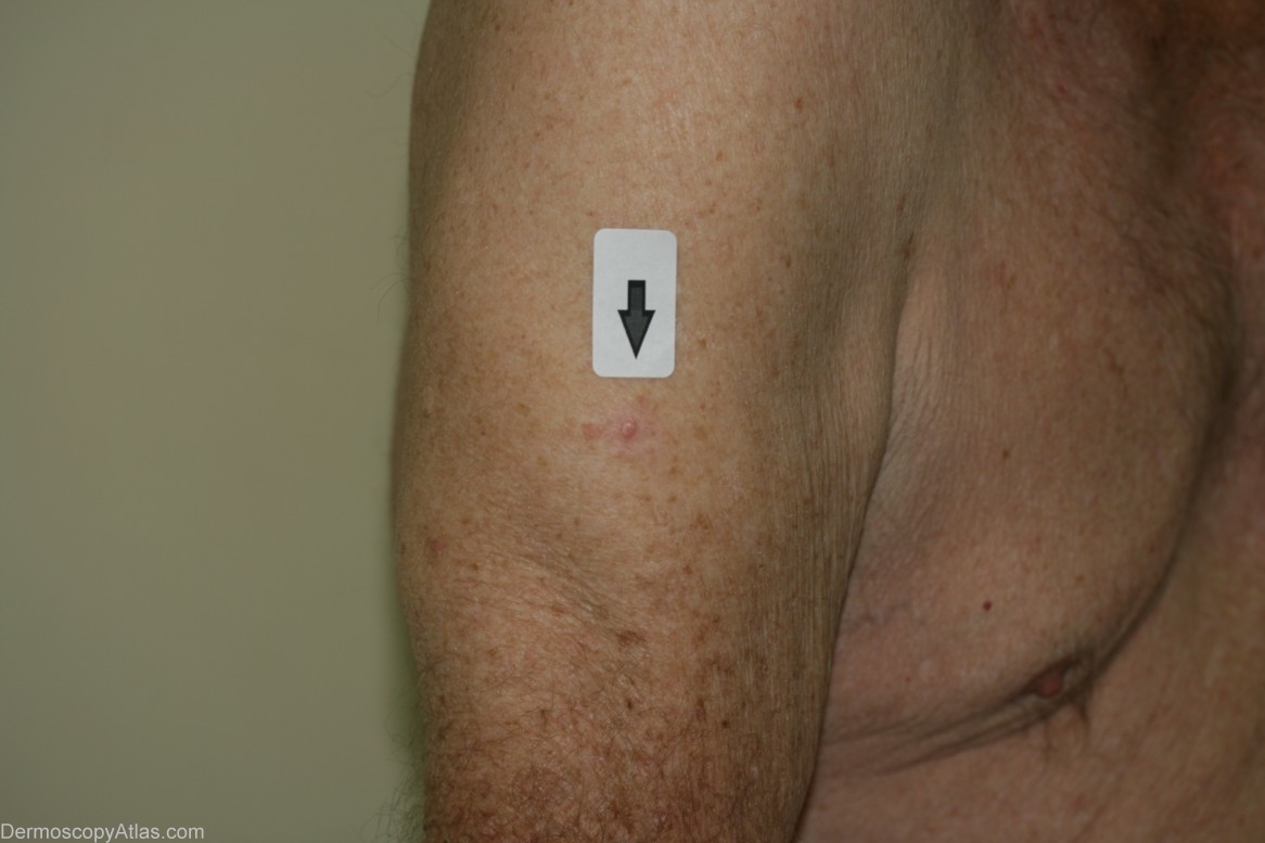
Site: Arm,upper
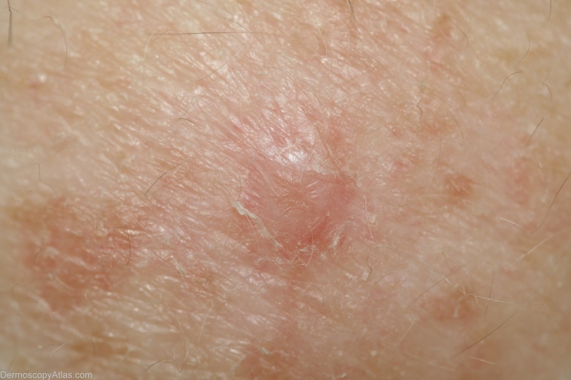
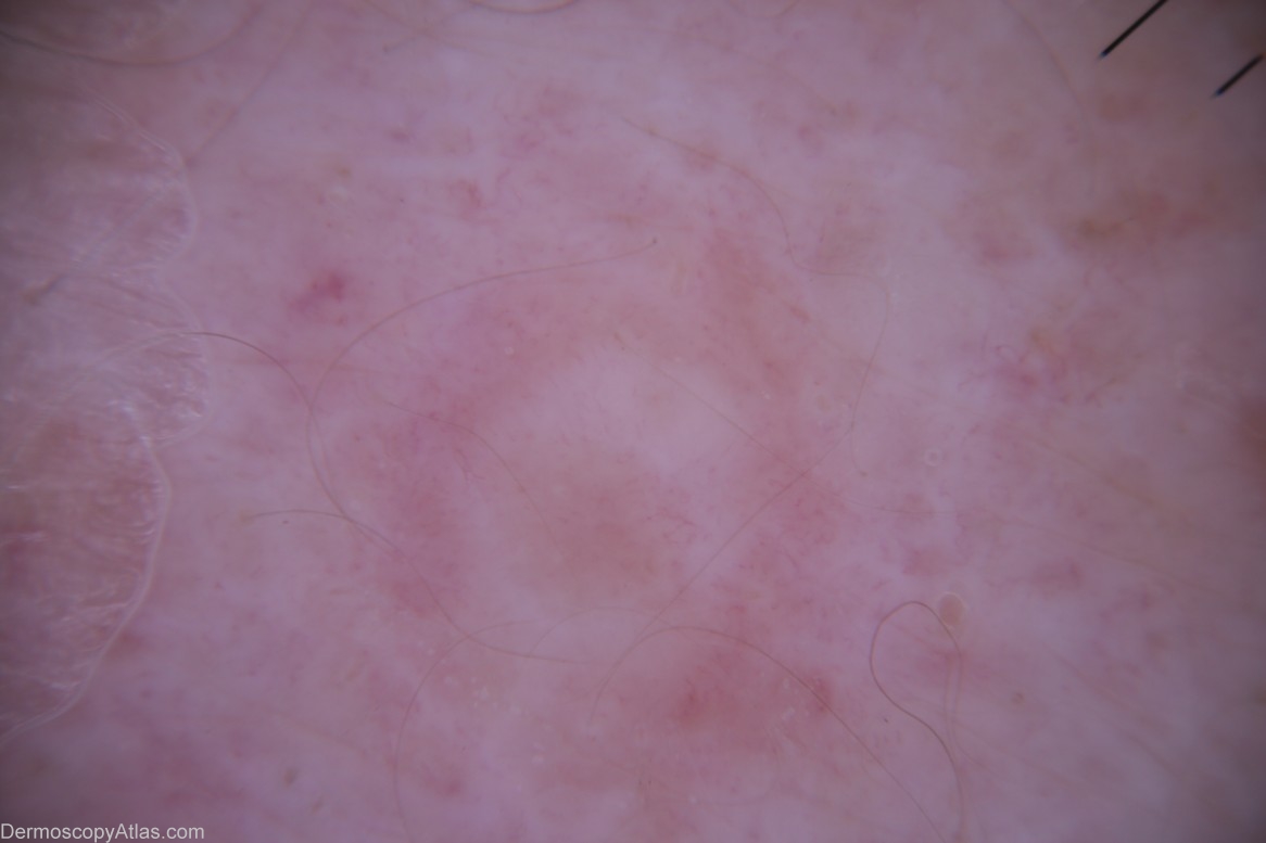
Diagnosis: Melanoma amelanotic
Sex: M
Age: 87
Type: Dermlite Non Polarised
Submitted By: Cliff Rosendahl
Description: Enhanced image (Dr Lester Cowell) to show "invisible" superficial spreading component
History: This 87 year old veteran of World War 2 had a regular skin examination ( Past history of non-melanoma skin cancers) and this small papule could not be confidently diagnosed clinically. It was thought to be benign but NMSC needed exclusion. It came back after a 4mm punch biopsy removal of the visible lesion as a level 3 amelanotic melanoma (Breslow thickness 1.3mm) with complete regression of the epidermal component. A further 1.5 cm clearance was obtained and the report came back as superficial spreading amelanotic melanoma (small residual level 2 0.3 mm thick)cleared by only 5mm. In retrospect, especially with enhancement of the image by Dr. Lester Cowell, the large insitu amelanotic melanoma can be visualised.